Explore
 Validate
 Learn
Learn Western blot
 Immunocytochemistry
Antibody data
- Antibody Data
- Antigen structure
- References [1]
- Comments [0]
- Validations
- Immunocytochemistry [1]
- Other assay [1]
Submit
Validation data
Reference
Comment
Report error
- Product number
- 701696 - Provider product page

- Provider
- Invitrogen Antibodies
- Product name
- BNIP3 Recombinant Rabbit Monoclonal Antibody (8H14L2)
- Antibody type
- Monoclonal
- Antigen
- Other
- Reactivity
- Human
- Host
- Rabbit
- Isotype
- IgG
- Antibody clone number
- 8H14L2
- Vial size
- 100 µg
- Concentration
- 0.5 mg/mL
- Storage
- Store at 4°C short term. For long term storage, store at -20°C, avoiding freeze/thaw cycles.
Submitted references Quantitative Ultrastructural Morphometry and Gene Expression of mTOR-Related Mitochondriogenesis within Glioblastoma Cells.
Ferese R, Lenzi P, Fulceri F, Biagioni F, Fabrizi C, Gambardella S, Familiari P, Frati A, Limanaqi F, Fornai F
International journal of molecular sciences 2020 Jun 27;21(13)
International journal of molecular sciences 2020 Jun 27;21(13)
No comments: Submit comment
Supportive validation
- Submitted by
- Invitrogen Antibodies (provider)
- Main image
- Experimental details
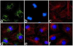
- Immunofluorescence was performed on methanol fixed Hela cells treated with Cobalt Chloride (15 µm/ 48 hours), for detection of BNIP3 using Anti-BNIP3 Recombinant Rabbit Monoclonal Antibody (Product # 701696, 1-2 µg/mL) and labeled with Goat anti-Rabbit IgG (H+L) Superclonal™ Secondary Antibody, Alexa Fluor® 488 conjugate (Product # A27034, 1:2000). Panel a) shows representative cells that were stained for detection and localization of BNIP3 protein (green), Panel b) is stained for nuclei (blue) using SlowFade® Gold Antifade Mountant with DAPI (Product # S36938). Panel c) represents cytoskeletal F-actin staining using Alexa Fluor® 555 Rhodamine Phalloidin (Product # R415, 1:300). Panel d) is a composite image of Panels a, b and c clearly demonstrating cytoplasmic localization of BNIP3. Panel e) represents merged image of untreated cells with no signal. Panel f) represents control cells with no primary Antibody to assess background.

Supportive validation
- Submitted by
- Invitrogen Antibodies (provider)
- Main image

- Experimental details
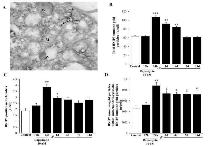
- Figure 18 Rapamycin increases the mitophagy marker BNIP3 at short-time intervals within U87MG cells. ( A ) Representative BNIP3-stained mitochondrion (indicated by the black arrow) at 24 h during 10 nM rapamycin, continuous exposure. Graphs report: ( B ) the amount of total BNIP3 particles; ( C ) the number of BNIP3-positive mitochondria; ( D ) the ratio of mitochondrial/cytosolic BNIP3 particles at various time interval during (12-24 h) or following (from 1 d up to 14 d) rapamycin exposure. M = mitochondrion, AV = vacuoles. * p