 Explore
 Validate
 Learn
Learn Western blot
Antibody data
- Antibody Data
- Antigen structure
- References [0]
- Comments [0]
- Validations
- Western blot [1]
- Immunocytochemistry [1]
Submit
Validation data
Reference
Comment
Report error
- Product number
- 710728 - Provider product page

- Provider
- Invitrogen Antibodies
- Product name
- BNIP3 Recombinant Polyclonal Antibody (8HCLC)
- Antibody type
- Polyclonal
- Antigen
- Other
- Reactivity
- Human
- Host
- Rabbit
- Isotype
- IgG
- Antibody clone number
- 8HCLC
- Vial size
- 100 µg
- Concentration
- 0.5 mg/mL
- Storage
- Store at 4°C short term. For long term storage, store at -20°C, avoiding freeze/thaw cycles.
No comments: Submit comment
Supportive validation
- Submitted by
- Invitrogen Antibodies (provider)
- Main image

- Experimental details
- Western blot analysis was performed on whole cell extracts (30 µg lysate) of HeLa (Lane 1), HeLa treated with Cobalt Chloride (150uM for 24 hours) (Lane 2). The blots were probed with Anti-BNIP3 Recombinant Rabbit Polyclonal Antibody (Product # 710728, 1-2 µg/mL) and detected by chemiluminescence using Goat anti-Rabbit IgG (H+L) Superclonal™ Secondary Antibody, HRP conjugate (Product # A27036, 0.4 µg/mL, 1:2500 dilution). The monomeric form of BNIP3 was dtetected as 30 kDa band was observed according to the treatment. Known quantity of protein samples were electrophoresed using Novex® NuPAGE® 12% Bis-Tris gel (Product # NP0342BOX), XCell SureLock™ Electrophoresis System (Product # EI0002) and Novex® Sharp Pre-Stained Protein Standard (Product # LC5800). Resolved proteins were then transferred onto a nitrocellulose membrane with iBlot® Dry Blotting System (Product # IB21001). The membrane was probed with the relevant primary and secondary Antibody following blocking with 5% skimmed milk. Chemiluminescent detection was performed using Pierce™ ECL Western blotting Substrate (Product # 32106).

Supportive validation
- Submitted by
- Invitrogen Antibodies (provider)
- Main image
- Experimental details
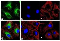
- For immunofluorescence analysis, cobalt chloride (150 uM, 48h) treated HeLa cells were fixed with methanol for detection of BNIP3 using Anti-BNIP3 Recombinant Rabbit Polyclonal Antibody (Product # 710728, 2 µg/mL) and labeled with Goat anti-Rabbit IgG (H+L) Superclonal Secondary Antibody, Alexa Fluor® 488 conjugate (Product # A27034, 1:2000). Cytoskeleton was stained with alpha-tubulin Monoclonal Antibody (Product # 32-2500, 1 µg/mL) followed by Goat anti-Mouse IgG Secondary Antibody, Alexa Fluor® 594 conjugate (Product # A-11032, 1:400). Panel a) shows representative cells that were stained for detection and localization of BNIP3 protein (green), Panel b) is stained for nuclei (blue) using SlowFade® Gold Antifade Mountant with DAPI (Product # S36938). Panel c) represents cytoskeletal alpha-tubulin staining (red). Panel d) is a composite image of Panels a, b and c clearly demonstrating cytoplasmic localization of BNIP3. Panel e) shows untreated cells with no signal. Panel f) represents control cells with no primary antibody to assess background.
