 Explore
 Validate
 Learn
Learn Western blot
Antibody data
- Antibody Data
- Antigen structure
- References [3]
- Comments [0]
- Validations
- Western blot [1]
Submit
Validation data
Reference
Comment
Report error
- Product number
- A01469 - Provider product page

- Provider
- Boster Biological Technology
- Product name
- Anti-BNIP3 Antibody Picoband™
- Antibody type
- Polyclonal
- Description
- Polyclonal antibody for BNIP3 detection. Host: Rabbit.Size: 100μg/vial. Tested applications: WB. Reactive species: Human. BNIP3 information: Subcellular Localization: Mitochondrion. Mitochondrion outer membrane; Coexpression with the EIB 19- kDa protein results in a shift in NIP3 localization pattern to the nuclear envelope. Colocalizes with ACAA2 in the mitochondria. Colocalizes with SPATA18 at the mitochondrion outer membrane.
- Reactivity
- Human, Mouse, Rat
- Host
- Rabbit
- Vial size
- 100μg/vial
- Concentration
- 0.5-1mg/ml, actual concentration vary by lot. Use suggested dilution ratio to decide dilution procedure.
- Storage
- At -20°C for one year. After reconstitution, at 4°C for one month. It can also be aliquoted and stored frozen at -20°C for a longer time. Avoid repeated freezing and thawing.
- Handling
- Add 0.2ml of distilled water will yield a concentration of 500ug/ml.
Submitted references Xiaojianzhong decoction prevents gastric precancerous lesions in rats by inhibiting autophagy and glycolysis in gastric mucosal cells.
β-arrestin1 inhibits hypoxic injury-induced autophagy in human pulmonary artery endothelial cells via the Akt/mTOR signaling pathway.
MicroRNA-145 induces apoptosis of glioma cells by targeting BNIP3 and Notch signaling.
Zhang JX, Bao SC, Chen J, Chen T, Wei HL, Zhou XY, Li JT, Yan SG
World journal of gastrointestinal oncology 2023 Mar 15;15(3):464-489
World journal of gastrointestinal oncology 2023 Mar 15;15(3):464-489
β-arrestin1 inhibits hypoxic injury-induced autophagy in human pulmonary artery endothelial cells via the Akt/mTOR signaling pathway.
Ning H, Deng J, Chen F, Liu Y, Kong D, Shan L, Zhang Z, Hu T
The international journal of biochemistry & cell biology 2020 Aug;125:105791
The international journal of biochemistry & cell biology 2020 Aug;125:105791
MicroRNA-145 induces apoptosis of glioma cells by targeting BNIP3 and Notch signaling.
Du Y, Li J, Xu T, Zhou DD, Zhang L, Wang X
Oncotarget 2017 Sep 22;8(37):61510-61527
Oncotarget 2017 Sep 22;8(37):61510-61527
No comments: Submit comment
Supportive validation
- Submitted by
- Boster Biological Technology (provider)
- Main image
- Experimental details
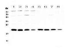
- Western blot analysis of BNIP3 using anti-BNIP3 antibody (A01469). Electrophoresis was performed on a 5-20% SDS-PAGE gel at 70V (Stacking gel) / 90V (Resolving gel) for 2-3 hours. The sample well of each lane was loaded with 50ug of sample under reducing conditions. Lane 1: rat brain tissue lysates,Lane 2: rat kidney tissue lysates,Lane 3: rat heart tissue lysates,Lane 4: rat testis tissue lysates,Lane 5: mouse brain tissue lysates,Lane 6: mouse kidney tissue lysates,Lane 7: mouse heart tissue lysates,Lane 8: mouse testis tissue lysates. After Electrophoresis, proteins were transferred to a Nitrocellulose membrane at 150mA for 50-90 minutes. Blocked the membrane with 5% Non-fat Milk/ TBS for 1.5 hour at RT. The membrane was incubated with rabbit anti-BNIP3 antigen affinity purified polyclonal antibody (Catalog # A01469) at 0.5 μg/mL overnight at 4°C, then washed with TBS-0.1%Tween 3 times with 5 minutes each and probed with a goat anti-rabbit IgG-HRP secondary antibody at a dilution of 1:10000 for 1.5 hour at RT. The signal is developed using an Enhanced Chemiluminescent detection (ECL) kit (Catalog # EK1002) with Tanon 5200 system. A specific band was detected for BNIP3 at approximately 21KD. The expected band size for BNIP3 is at 21KD.
- Additional image


