Explore
 Validate
 Learn
Learn Immunocytochemistry
Antibody data
- Antibody Data
- Antigen structure
- References [2]
- Comments [0]
- Validations
- Immunocytochemistry [3]
- Immunohistochemistry [1]
- Other assay [1]
Submit
Validation data
Reference
Comment
Report error
- Product number
- PA5-11402 - Provider product page

- Provider
- Invitrogen Antibodies
- Product name
- BNIP3 Polyclonal Antibody
- Antibody type
- Polyclonal
- Antigen
- Synthetic peptide
- Reactivity
- Human, Mouse
- Host
- Rabbit
- Isotype
- IgG
- Vial size
- 400 μL
- Concentration
- 1.2 mg/mL
- Storage
- Store at 4°C short term. For long term storage, store at -20°C, avoiding freeze/thaw cycles.
Submitted references Blockade of the CD93 pathway normalizes tumor vasculature to facilitate drug delivery and immunotherapy.
Proximity-dependent initiation of hybridization chain reaction.
Sun Y, Chen W, Torphy RJ, Yao S, Zhu G, Lin R, Lugano R, Miller EN, Fujiwara Y, Bian L, Zheng L, Anand S, Gao F, Zhang W, Ferrara SE, Goodspeed AE, Dimberg A, Wang XJ, Edil BH, Barnett CC, Schulick RD, Chen L, Zhu Y
Science translational medicine 2021 Jul 28;13(604)
Science translational medicine 2021 Jul 28;13(604)
Proximity-dependent initiation of hybridization chain reaction.
Koos B, Cane G, Grannas K, Löf L, Arngården L, Heldin J, Clausson CM, Klaesson A, Hirvonen MK, de Oliveira FM, Talibov VO, Pham NT, Auer M, Danielson UH, Haybaeck J, Kamali-Moghaddam M, Söderberg O
Nature communications 2015 Jun 12;6:7294
Nature communications 2015 Jun 12;6:7294
No comments: Submit comment
Supportive validation
- Submitted by
- Invitrogen Antibodies (provider)
- Main image

- Experimental details
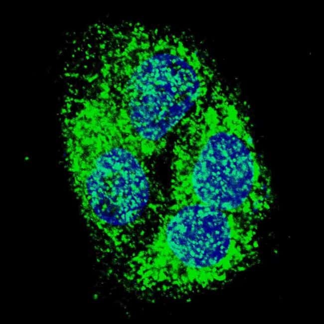
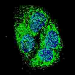
- Immunofluorescent analysis of HepG2 cells using a BNIP3 polyclonal antibody (Product # PA5-11402). HepG2 cells were fixed with 4% PFA (20 min), permeabilized with Triton X-100 (0.2%, 30 min). Cells were then incubated with a BNIP3 polyclonal antibody (Product # PA5-11402) (1:500, 2 hr at room temperature). Primary antibody detected by fluor-conjugated donkey anti-rabbit secondary antibody (green) was used (1:1000, 1hr). Nuclei were counterstained with Hoechst 33342 (blue) (10 µg/mL, 5 min).
- Submitted by
- Invitrogen Antibodies (provider)
- Main image

- Experimental details
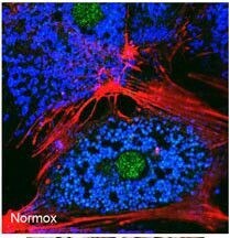
- Immunofluorescent analysis of mouse hepatocytes using a BNIP3 polyclonal antibody (Product # PA5-11402) at a dilution of 1:50. Freshly isolated mouse hepatocytes plated on coverslips (2x10^5 cells/22-mm glass coverslip) were cultured under normoxic conditions for 6 hr. The cells were then fixed in 2% paraformaldehyde in PBS for 1 hr, and processed for confocal immunofluorescence (red: F-actin, blue: ATP-synthase, green: BNIP3).
- Submitted by
- Invitrogen Antibodies (provider)
- Main image
- Experimental details
- Immunocytochemistry analysis of BNIP3 in HepG2 cells. Samples were incubated with BNIP3 polyclonal antibody (Product # PA5-11402) using a dilution of 1:500 for 2 h at room temperature followed by Alexa Fluor® 488 conjugated donkey anti-rabbit at a dilution of 1:1,000 for 1hr. Cells were fixed with 4% PFA (20 min) and permeabilized with Triton X-100 (0.2%, 30 min). Nuclei were counterstained with Hoechst 33342 (blue) (10 μg/mL, 5 min). BNIP3 immunoreactivity is localized to the cytoplasm of HepG2 cells.

Supportive validation
- Submitted by
- Invitrogen Antibodies (provider)
- Main image
- Experimental details
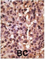
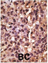
- Immunohistochemistry analysis of BNIP3 in formalin-fixed and paraffin-embedded human cancer tissue. Samples were incubated with BNIP3 polyclonal antibody (Product # PA5-11402) which was peroxidase-conjugated to the secondary antibody, followed by DAB staining. This data demonstrates the use of this antibody for immunohistochemistry; clinical relevance has not been evaluated. BC = breast carcinoma; HC = hepatocarcinoma.
Supportive validation
- Submitted by
- Invitrogen Antibodies (provider)
- Main image
- Experimental details
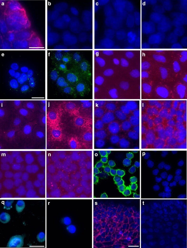
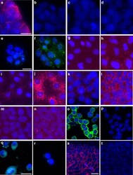
- Figure 4 In situ prox-HCR. ( a-d ) Technical controls for the E-cadherin/beta-catenin interaction. Strong membranous signal could be observed in HT29 cells when both primary antibodies were applied ( a ), while omitting either one of the primary antibodies ( b , c ) or both ( d ) did not yield visible signal. Phosphorylation of platelet-derived growth factor receptor-beta (PDGFR-beta) could be shown in BjHTert cells following stimulation with 100 ng ml -1 ( f ), while expression of phosphorylated receptor was low in non-stimulated cells ( e ). ProxHCR was used to visualize the induction of autophagy following starvation and incubation with CoCl 2 in Caco cells ( g-j ). Although untreated cells showed only low basal activity ( g ) of BCL2-BNIP3 interaction, a highly increased signal could be observed in treated cells ( h ). The same holds true for LC3-SQSTM1 interaction ( i , j ). The MEK-ERK interaction could be induced in A431 cells by stimulation with 10 ng ml -1 EGF for 10 min ( l ), while the non-stimulated cells only showed low basal signal ( k ). Under the same conditions phosphorylation of Akt could be observed ( n ), while no phosphorylation was visible in non-stimulated cells ( m ). In panel o , the phosphorylation of Syk is shown ( p : no primary antibodies). Detection of Her2 protein was possible as well and shown in panel q ( r : no primary antibodies). Expression of E-cadherin/beta-catenin interaction could be observed even in FFPE skin tissue ( s ), while no sig