Explore
 Validate
 Learn
Learn Western blot
Antibody data
- Antibody Data
- Antigen structure
- References [4]
- Comments [0]
- Validations
- Western blot [1]
- Immunohistochemistry [1]
Submit
Validation data
Reference
Comment
Report error
- Product number
- sc-20812 - Provider product page

- Provider
- Santa Cruz Biotechnology
- Proper citation
- Santa Cruz Biotechnology Cat#sc-20812, RRID:AB_2274338
- Product name
- Anti-AQP4
- Antibody type
- Polyclonal
- Antigen
- Recombinant full-length protein
- Reactivity
- Human
- Host
- Rabbit
Submitted references Glial dystrophin-associated proteins, laminin and agrin, are downregulated in the brain of mdx mouse
Transcriptional activation of endothelial cells by TGFβ coincides with acute microvascular plasticity following focal spinal cord ischaemia/reperfusion injury.
Altered regulation of aquaporin gene expression in allergen and IL-13-induced mouse models of asthma.
Close association of water channel AQP1 with amyloid-beta deposition in Alzheimer disease brains.
Beatrice Nico, Roberto Tamma, Tiziana Annese, Domenica Mangieri, Annamaria De Luca, Patrizia Corsi, Vincenzo Benagiano, Vito Longo, Enrico Crivellato, Andrea Salmaggi, Domenico Ribatti
Laboratory Investigation 2010 Aug;90(11):1645-1660
Laboratory Investigation 2010 Aug;90(11):1645-1660
Transcriptional activation of endothelial cells by TGFβ coincides with acute microvascular plasticity following focal spinal cord ischaemia/reperfusion injury.
Benton RL, Maddie MA, Dincman TA, Hagg T, Whittemore SR
ASN neuro 2009 Aug 26;1(3)
ASN neuro 2009 Aug 26;1(3)
Altered regulation of aquaporin gene expression in allergen and IL-13-induced mouse models of asthma.
Krane CM, Deng B, Mutyam V, McDonald CA, Pazdziorko S, Mason L, Goldman S, Kasaian M, Chaudhary D, Williams C, Ho MW
Cytokine 2009 Apr;46(1):111-8
Cytokine 2009 Apr;46(1):111-8
Close association of water channel AQP1 with amyloid-beta deposition in Alzheimer disease brains.
Misawa T, Arima K, Mizusawa H, Satoh J
Acta neuropathologica 2008 Sep;116(3):247-60
Acta neuropathologica 2008 Sep;116(3):247-60
No comments: Submit comment
Supportive validation
- Submitted by
- per
- Main image
- Experimental details
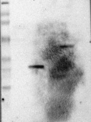
- Western blot analysis of antibody specificity using a routine panel composed of IgG/HSA-depleted human plasma and protein lysates from selected human tissues and cell lines.
- Validation comment
- Band of predicted size in kDa (+/-20%) with additional bands present.
- Primary Ab dilution
- 1:500
- Secondary Ab dilution
- 1:3000
- Lane 1
- Marker [kDa]: 230, 110, 82, 49.3, 32.2, 25.5, 17.6
- Lane 2
- RT-4
- Lane 3
- U-251MG sp
- Lane 4
- Human Plasma
- Lane 5
- Liver
- Lane 6
- Tonsil
- Theoretical target weight
- [kDa] 35

Supportive validation
- Submitted by
- per
- Main image
- Experimental details
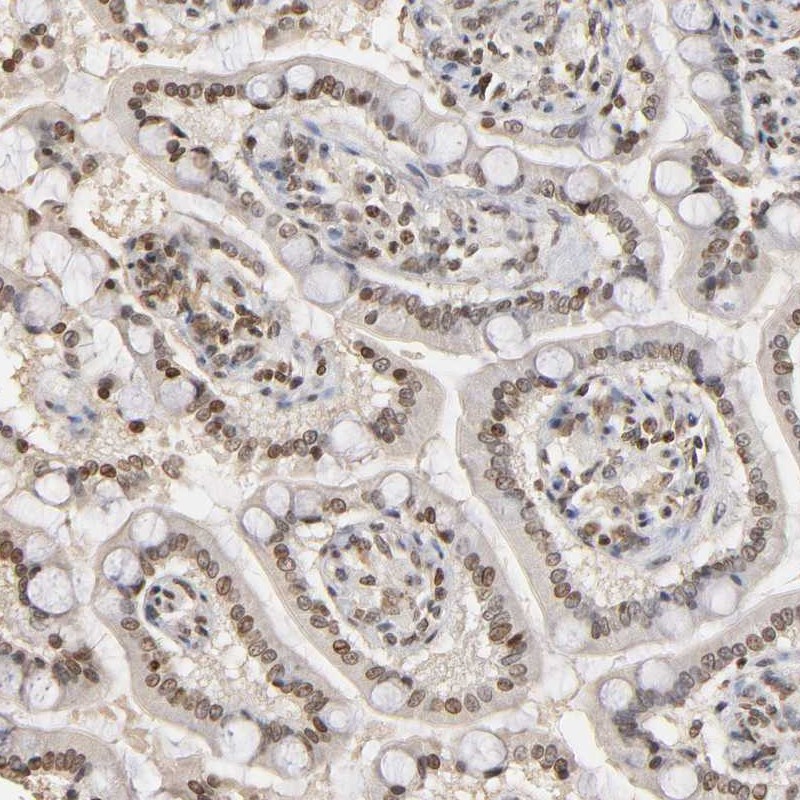
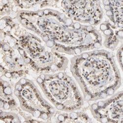
- Immunohistochemical staining of human small intestine shows moderate nuclear positivity in glandular cells.
- Validation comment
- Staining pattern partly consistent with experimental and/or bioinformatic data.