Explore
 Validate
 Learn
Learn Western blot
 Immunohistochemistry
Antibody data
- Antibody Data
- Antigen structure
- References [6]
- Comments [0]
- Validations
- Western blot [1]
Submit
Validation data
Reference
Comment
Report error
- Product number
- PB9475 - Provider product page

- Provider
- Boster Biological Technology
- Product name
- Anti-Aquaporin 4/AQP4 Antibody Picoband™
- Antibody type
- Polyclonal
- Description
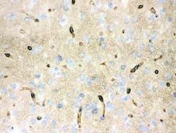
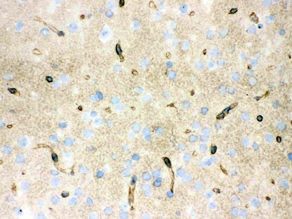
- Polyclonal antibody for AQUAPORIN 4/AQP4 detection. Host: Rabbit.Size: 100μg/vial. Tested applications: IHC-P. Reactive species: Human. AQUAPORIN 4/AQP4 information: Molecular Weight: 34830 MW; Subcellular Localization: Membrane; Multi-pass membrane protein; Tissue Specificity: Brain - muscle >> heart, kidney, lung, and trachea.
- Reactivity
- Human, Mouse, Rat
- Host
- Rabbit
- Vial size
- 100μg/vial
- Concentration
- Add 0.2ml of distilled water will yield a concentration of 500ug/ml.
- Storage
- At -20°C for one year. After reconstitution, at 4°C for one month. It can also be aliquoted and stored frozen at -20°C for a longer time. Avoid repeated freezing and thawing.
- Handling
- Add 0.2ml of distilled water will yield a concentration of 500ug/ml.
Submitted references Long-term impact of maternal obesity on the gliovascular unit and ephrin signaling in the hippocampus of adult offspring.
Proteomics identifies lipocalin-2 in neonatal inflammation associated with cerebrovascular alteration in mice and preterm infants.
Sex-Dependent Gliovascular Interface Abnormality in the Hippocampus following Postnatal Immune Activation in Mice.
Group 2 innate lymphoid cells suppress the pathology of neuromyelitis optica spectrum disorder.
Hippocampal Atrophy Following Subarachnoid Hemorrhage Correlates with Disruption of Astrocyte Morphology and Capillary Coverage by AQP4.
Brain edema after intracerebral hemorrhage in rats: the role of iron overload and aquaporin 4.
Shiadeh SMJ, Goretta F, Svedin P, Jansson T, Mallard C, Ardalan M
Journal of neuroinflammation 2024 Feb 2;21(1):39
Journal of neuroinflammation 2024 Feb 2;21(1):39
Proteomics identifies lipocalin-2 in neonatal inflammation associated with cerebrovascular alteration in mice and preterm infants.
Gravina G, Ardalan M, Chumak T, Nilsson AK, Ek JC, Danielsson H, Svedin P, Pekny M, Pekna M, Sävman K, Hellström A, Mallard C
iScience 2023 Jul 21;26(7):107217
iScience 2023 Jul 21;26(7):107217
Sex-Dependent Gliovascular Interface Abnormality in the Hippocampus following Postnatal Immune Activation in Mice.
Ardalan M, Chumak T, Quist A, Jabbari Shiadeh SM, Mallard AJ, Rafati AH, Mallard C
Developmental neuroscience 2022;44(4-5):320-330
Developmental neuroscience 2022;44(4-5):320-330
Group 2 innate lymphoid cells suppress the pathology of neuromyelitis optica spectrum disorder.
Kong Y, Li HD, Wang D, Gao X, Yang C, Li M, Chang T, Liu Q
FASEB journal : official publication of the Federation of American Societies for Experimental Biology 2021 Nov;35(11):e21856
FASEB journal : official publication of the Federation of American Societies for Experimental Biology 2021 Nov;35(11):e21856
Hippocampal Atrophy Following Subarachnoid Hemorrhage Correlates with Disruption of Astrocyte Morphology and Capillary Coverage by AQP4.
Anzabi M, Ardalan M, Iversen NK, Rafati AH, Hansen B, Østergaard L
Frontiers in cellular neuroscience 2018;12:19
Frontiers in cellular neuroscience 2018;12:19
Brain edema after intracerebral hemorrhage in rats: the role of iron overload and aquaporin 4.
Qing WG, Dong YQ, Ping TQ, Lai LG, Fang LD, Min HW, Xia L, Heng PY
Journal of neurosurgery 2009 Mar;110(3):462-8
Journal of neurosurgery 2009 Mar;110(3):462-8
No comments: Submit comment
Supportive validation
- Submitted by
- Boster Biological Technology (provider)
- Main image
- Experimental details
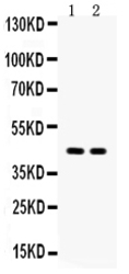
- Western blot analysis of Aquaporin 4 using anti-Aquaporin 4 antibody (PB9475). Electrophoresis was performed on a 5-20% SDS-PAGE gel at 70V (Stacking gel) / 90V (Resolving gel) for 2-3 hours. The sample well of each lane was loaded with 50ug of sample under reducing conditions. Lane 1: Rat Brain Tissue Lysate, Lane 2: Mouse Brain Tissue Lysate. After Electrophoresis, proteins were transferred to a Nitrocellulose membrane at 150mA for 50-90 minutes. Blocked the membrane with 5% Non-fat Milk/ TBS for 1.5 hour at RT. The membrane was incubated with rabbit anti-Aquaporin 4 antigen affinity purified polyclonal antibody (Catalog # PB9475) at 0.5 μg/mL overnight at 4°C, then washed with TBS-0.1%Tween 3 times with 5 minutes each and probed with a goat anti-rabbit IgG-HRP secondary antibody at a dilution of 1:10000 for 1.5 hour at RT. The signal is developed using an Enhanced Chemiluminescent detection (ECL) kit (Catalog # EK1002) with Tanon 5200 system. A specific band was detected for Aquaporin 4 at approximately 45KD. The expected band size for Aquaporin 4 is at 35KD.
- Additional image