Explore
 Validate
 Learn
Learn Western blot
 ELISA
ELISAAntibody data
- Antibody Data
- Antigen structure
- References [4]
- Comments [0]
- Validations
- Western blot [2]
- Flow cytometry [2]
Submit
Validation data
Reference
Comment
Report error
- Product number
- NBP1-30249 - Provider product page

- Provider
- Novus Biologicals
- Proper citation
- Novus Cat#NBP1-30249, RRID:AB_1987289
- Product name
- Mouse Monoclonal Myosin Light Chain 2 Antibody
- Antibody type
- Monoclonal
- Description
- Protein G purified.
- Reactivity
- Human, Mouse, Xenopus
- Host
- Mouse
- Isotype
- IgG
- Vial size
- 0.1 ml
- Concentration
- 1.0 mg/ml
- Storage
- Store at 4C short term. Aliquot and store at -20C long term. Avoid freeze-thaw cycles.
Submitted references Binary Colloidal Crystals Drive Spheroid Formation and Accelerate Maturation of Human-Induced Pluripotent Stem Cell-Derived Cardiomyocytes.
ULK1 prevents cardiac dysfunction in obesity through autophagy-meditated regulation of lipid metabolism.
Early ketamine exposure results in cardiac enlargement and heart dysfunction in Xenopus embryos.
The nuclear F-actin interactome of Xenopus oocytes reveals an actin-bundling kinesin that is essential for meiotic cytokinesis.
Cui C, Wang J, Qian D, Huang J, Lin J, Kingshott P, Wang PY, Chen M
ACS applied materials & interfaces 2019 Jan 30;11(4):3679-3689
ACS applied materials & interfaces 2019 Jan 30;11(4):3679-3689
ULK1 prevents cardiac dysfunction in obesity through autophagy-meditated regulation of lipid metabolism.
An M, Ryu DR, Won Park J, Ha Choi J, Park EM, Eun Lee K, Woo M, Kim M
Cardiovascular research 2017 Aug 1;113(10):1137-1147
Cardiovascular research 2017 Aug 1;113(10):1137-1147
Early ketamine exposure results in cardiac enlargement and heart dysfunction in Xenopus embryos.
Guo R, Liu G, Du M, Shi Y, Jiang P, Liu X, Liu L, Liu J, Xu Y
BMC anesthesiology 2016 Apr 18;16:23
BMC anesthesiology 2016 Apr 18;16:23
The nuclear F-actin interactome of Xenopus oocytes reveals an actin-bundling kinesin that is essential for meiotic cytokinesis.
Samwer M, Dehne HJ, Spira F, Kollmar M, Gerlich DW, Urlaub H, Görlich D
The EMBO journal 2013 Jul 3;32(13):1886-902
The EMBO journal 2013 Jul 3;32(13):1886-902
No comments: Submit comment
Supportive validation
- Submitted by
- Novus Biologicals (provider)
- Main image

- Experimental details
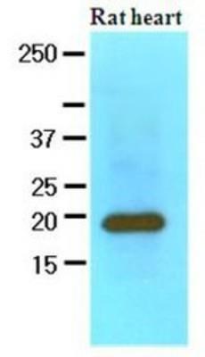
- Western Blot: Myosin Light Chain 2 Antibody (3B2) [NBP1-30249] - The extracts of Rat heart (60ug) were resolved by SDS-PAGE, transferred to NC membrane and probed with anti-human MYL2 (1:1000). Proteins were visualized using a goat anti-mouse secondary antibody conjugated to HRP and an ECL detection system.
- Submitted by
- Novus Biologicals (provider)
- Main image
- Experimental details
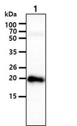
- Western Blot: Myosin Light Chain 2 Antibody (3B2) [NBP1-30249] - The tissue lysate (40ug) was resolved by SDS-PAGE, transferred to PVDF membrane and probed with anti-human MYL2 antibody (1:1000). Proteins were visualized using a goat anti-mouse secondary antibody conjugated to HRP and an ECL detection system. Lane 1.: Mouse heart tissue lysate
Supportive validation
- Submitted by
- Novus Biologicals (provider)
- Main image
- Experimental details
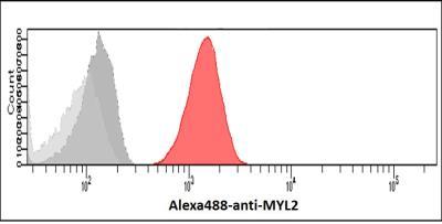
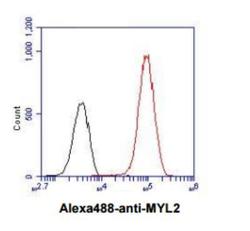
- Flow Cytometry: Myosin Light Chain 2 Antibody (3B2) [NBP1-30249] - Flow cytometry analysis of MYL2 in A431 cell line, staining at 2-5ug for 1x10⁶cells (red line). The secondary antibody used goat anti-mouse IgG Alexa fluor 488 conjugate. Isotype control antibody was mouse IgG (black line).
- Submitted by
- Novus Biologicals (provider)
- Main image

- Experimental details
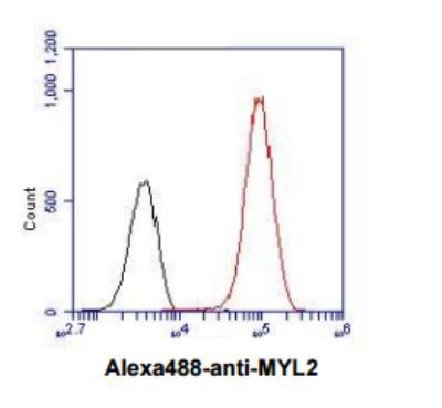
- Flow Cytometry: Myosin Light Chain 2 Antibody (3B2) [NBP1-30249] - Analysis of MYL2 in A431 cells. The cell was stained at 2-5ug for 1x10^6cells (red). A Goat anti mouse IgG (Alexa fluor 488) was used as the secondary antibody. Mouse monoclonal IgG was used as the isotype control (dark gray), cells without incubation with primary and secondary antibody was used as the negative control (light gray).