Explore
 Validate
 Learn
Learn Western blot
Antibody data
- Antibody Data
- Antigen structure
- References [0]
- Comments [0]
- Validations
- Western blot [3]
- Immunocytochemistry [2]
- Immunohistochemistry [1]
- Flow cytometry [2]
Submit
Validation data
Reference
Comment
Report error
- Product number
- ALM-051-100UG - Provider product page

- Provider
- Invitrogen Antibodies
- Product name
- KCNN4 (KCa3.1, SK4) (extracellular) Monoclonal Antibody (6C1)
- Antibody type
- Monoclonal
- Antigen
- Other
- Reactivity
- Human, Mouse, Rat
- Host
- Mouse
- Isotype
- IgM
- Antibody clone number
- 6C1
- Vial size
- 100 µg
- Concentration
- 1 mg/mL
- Storage
- -20° C, Avoid Freeze/Thaw Cycles
No comments: Submit comment
Supportive validation
- Submitted by
- Invitrogen Antibodies (provider)
- Main image

- Experimental details
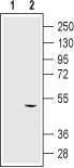
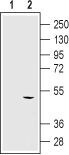
- Western blot analysis using Mouse Anti-KCNN4 (KCa3.1, SK4) (extracellular) Antibody (#ALM-051), (1:250): - 1. HEK cells transfected with control vector. 2. HEK cells transfected with human KCa3.1.
- Submitted by
- Invitrogen Antibodies (provider)
- Main image
- Experimental details
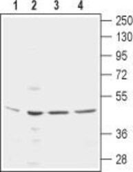
- Western blot analysis using Mouse Anti-KCNN4 (KCa3.1, SK4) (extracellular) Antibody (#ALM-051), (1:250): - 1. Mouse MS1 endothelial cells. 2. Rat IEC-6 intestinal epithelial cells. 3. Human LN-CaP prostate carcinoma cells. 4. Human THP-1 acute monocytic leukemia cells.
- Submitted by
- Invitrogen Antibodies (provider)
- Main image

- Experimental details
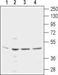
- Western blot analysis using Mouse Anti-KCNN4 (KCa3.1, SK4) (extracellular) Antibody (#ALM-051), (1:250): - 1. HEK cells transfected with control vector. 2. HEK cells transfected with human KCa3.1.
Supportive validation
- Submitted by
- Invitrogen Antibodies (provider)
- Main image
- Experimental details
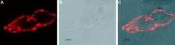
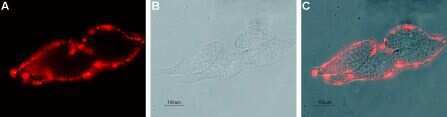
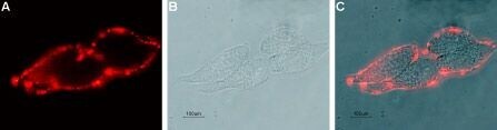
- Expression of KCNN4in live intact human LN-CaP prostate carcinoma cells - Cell surface detection of KCNN4 in live intact human LN-CaP prostate carcinoma cells with Mouse Anti-KCNN4 (KCa3.1, SK4) (extracellular) Antibody (#ALM-051) (1:20), followed by goat- Anti-mouse-DyLight-594 secondary Antibody (red) (A). B. Live view of the cells. C. Merge of the two images.
- Submitted by
- Invitrogen Antibodies (provider)
- Main image

- Experimental details
- Expression of KCNN4in live intact human LN-CaP prostate carcinoma cells - Cell surface detection of KCNN4 in live intact human LN-CaP prostate carcinoma cells with Mouse Anti-KCNN4 (KCa3.1, SK4) (extracellular) Antibody (#ALM-051) (1:20), followed by goat- Anti-mouse-DyLight-594 secondary Antibody (red) (A). B. Live view of the cells. C. Merge of the two images.
Supportive validation
- Submitted by
- Invitrogen Antibodies (provider)
- Main image
- Experimental details
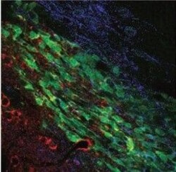
- Multiplex staining of KCa3.1 (SK4) and TRPC1 in mouse brain - Immunohistochemical staining of mouse brain sections using Mouse Anti-KCNN4 (KCa3.1, SK4) (extracellular) Antibody (#ALM-051) and Anti-TRPC1 Antibody (#ACC-010). TRPC1 staining (blue) is detected in neuroblasts and outside neuroblasts as well (in astrocytes). KCa3.1 (red) is detected in neuroblasts. Merged image demonstrates the partial co-localization between the two. Adapted from Turner, K.L.et al. (2014)Cereb. Cortex.24,2388. with permissionof Oxford University Press.

Supportive validation
- Submitted by
- Invitrogen Antibodies (provider)
- Main image

- Experimental details
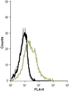
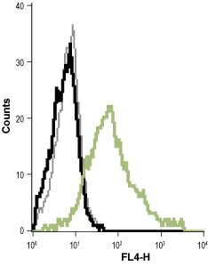
- Cell surface detection of KCa3.1 (SK4) in live intact THP-1 (human acute monocytic leukemia cells) cell line: - (black) Cells + Goat- Anti-mouse-Cy5. (red) Cells + Mouse IgM isotype control+ Goat- Anti-mouse-Cy5. (green) Cells + Mouse Anti-KCNN4 (KCa3.1, SK4) (extracellular) Antibody (#ALM-051), (1:20) + Goat- Anti-mouse-Cy5.
- Submitted by
- Invitrogen Antibodies (provider)
- Main image
- Experimental details
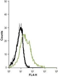
- Cell surface detection of KCa3.1 (SK4) in live intact Raji (human Burkitts lymphoma B cells) cell line: - (black) Cells + goat- Anti-mouse-Cy5. (red) Cells + mouse IgM isotype control + goat- Anti-mouse-Cy5. (green) Cells + Mouse Anti-KCNN4 (KCa3.1, SK4) (extracellular) Antibody (#ALM-051), (1:20) + goat- Anti-mouse-Cy5.