 Explore
 Validate
 Learn
Learn Western blot
 Immunocytochemistry
Antibody data
- Antibody Data
- Antigen structure
- References [1]
- Comments [0]
- Validations
- Immunocytochemistry [3]
- Immunohistochemistry [1]
- Other assay [1]
Submit
Validation data
Reference
Comment
Report error
- Product number
- PA5-27545 - Provider product page

- Provider
- Invitrogen Antibodies
- Product name
- RPL29 Polyclonal Antibody
- Antibody type
- Polyclonal
- Antigen
- Recombinant full-length protein
- Description
- Recommended positive controls: NT2D1, PC-3, U87-MG, SK-N-SH. Predicted reactivity: Mouse (84%), Rat (85%), Pig (93%), Rhesus Monkey (97%), Chimpanzee (96%), Bovine (93%). Store product as a concentrated solution. Centrifuge briefly prior to opening the vial.
- Reactivity
- Human
- Host
- Rabbit
- Isotype
- IgG
- Vial size
- 100 μL
- Concentration
- 1 mg/mL
- Storage
- Store at 4°C short term. For long term storage, store at -20°C, avoiding freeze/thaw cycles.
Submitted references Knockdown of the Ribosomal Protein eL29 in Mammalian Cells Leads to Significant Changes in Gene Expression at the Transcription Level.
Gopanenko AV, Kolobova AV, Meschaninova MI, Venyaminova AG, Tupikin AE, Kabilov MR, Malygin AA, Karpova GG
Cells 2020 May 15;9(5)
Cells 2020 May 15;9(5)
No comments: Submit comment
Supportive validation
- Submitted by
- Invitrogen Antibodies (provider)
- Main image
- Experimental details
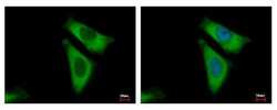
- RPL29 Polyclonal Antibody detects RPL29 protein at cytoplasm by immunofluorescent analysis. Sample: HeLa cells were fixed in 4% paraformaldehyde at RT for 15 min. Green: RPL29 protein stained by RPL29 Polyclonal Antibody (Product # PA5-27545) diluted at 1:500. Blue: Hoechst 33343 staining.

- Submitted by
- Invitrogen Antibodies (provider)
- Main image
- Experimental details
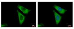
- RPL29 Polyclonal Antibody detects RPL29 protein at cytoplasm by immunofluorescent analysis. Sample: HeLa cells were fixed in 4% paraformaldehyde at RT for 15 min. Green: RPL29 protein stained by RPL29 Polyclonal Antibody (Product # PA5-27545) diluted at 1:500. Blue: Hoechst 33343 staining.

- Submitted by
- Invitrogen Antibodies (provider)
- Main image

- Experimental details
- RPL29 Polyclonal Antibody detects RPL29 protein at cytoplasm by immunofluorescent analysis. Sample: HeLa cells were fixed in 4% paraformaldehyde at RT for 15 min. Green: RPL29 protein stained by RPL29 Polyclonal Antibody (Product # PA5-27545) diluted at 1:500. Blue: Hoechst 33343 staining.

Supportive validation
- Submitted by
- Invitrogen Antibodies (provider)
- Main image

- Experimental details
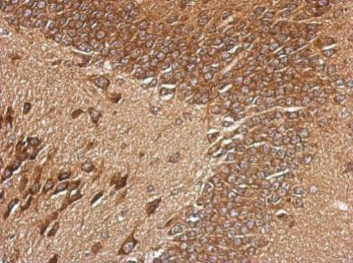
- Immunohistochemical analysis of paraffin-embedded CL1-5 xenograft, using RPL29 (Product # PA5-27545) antibody at 1:500 dilution. Antigen Retrieval: EDTA based buffer, pH 8.0, 15 min.
Supportive validation
- Submitted by
- Invitrogen Antibodies (provider)
- Main image

- Experimental details
- Figure 1 Characterization of HEK293 cells knocked down of ribosomal protein eL29. ( A ) Western blot analysis of the levels of ribosomal proteins eL29 and uS2 (as a reference) in cells transfected with siRNAs specific for eL29 mRNA with scrambled control, 1 and 2 days after transfection. The diagrams show the western blot data in triplicate as the mean of arbitrary units (a.u.) +- CEM (** p < 0.01, calculated using Student's t-test). ( B ) Histograms of MTT assay of cells transfected with scrambled or eL29 mRNA-specific siRNAs. ( C ) Electrophoretic analysis of total RNA samples extracted from cells transfected with the above siRNAs on a 0.5% agarose gel stained with ethidium bromide. ( D ) Polysome profiles obtained by centrifugation of lysates of cells transfected with scrambled (solid line) and eL29 mRNA-specific (dashed line) siRNAs in sucrose density gradient with marked peaks corresponding to 60S subunits, 80S ribosomes and polysomes. Western-blot analysis of gradient fractions for the presence of eL29 and uL18 (as a reference). Lanes 1, 2 and 3 correspond to the sucrose gradient fractions containing ""heavy"" polysomes (>5 ribosomes per mRNA), ""light"" polysomes (
