 Explore
 Validate
 Learn
Learn Western blot
Antibody data
- Antibody Data
- Antigen structure
- References [2]
- Comments [0]
- Validations
- Western blot [1]
- Immunocytochemistry [1]
Submit
Validation data
Reference
Comment
Report error
- Product number
- AF3364 - Provider product page

- Provider
- R&D Systems
- Product name
- Human Pax2 Antibody
- Antibody type
- Polyclonal
- Description
- Antigen Affinity-purified. Detects human Pax2 in direct ELISAs and Western blots. In direct ELISAs, less than 10% cross-reactivity with recombinant human Pax5 is observed.
- Reactivity
- Human
- Host
- Goat
- Conjugate
- Unconjugated
- Antigen sequence
Q02962- Isotype
- IgG
- Vial size
- 100 ug
- Concentration
- LYOPH
- Storage
- Use a manual defrost freezer and avoid repeated freeze-thaw cycles. 12 months from date of receipt, -20 to -70 °C as supplied. 1 month, 2 to 8 °C under sterile conditions after reconstitution. 6 months, -20 to -70 °C under sterile conditions after reconstitution.
Submitted references Bcl-2 Expression in Pericytes and Astrocytes Impacts Vascular Development and Homeostasis.
Histone deacetylase expression patterns in developing murine optic nerve.
Zaitoun IS, Wintheiser CM, Jamali N, Wang S, Suscha A, Darjatmoko SR, Schleck K, Hanna BA, Lindner V, Sheibani N, Sorenson CM
Scientific reports 2019 Jul 4;9(1):9700
Scientific reports 2019 Jul 4;9(1):9700
Histone deacetylase expression patterns in developing murine optic nerve.
Tiwari S, Dharmarajan S, Shivanna M, Otteson DC, Belecky-Adams TL
BMC developmental biology 2014 Jul 9;14:30
BMC developmental biology 2014 Jul 9;14:30
No comments: Submit comment
Supportive validation
- Submitted by
- R&D Systems (provider)
- Main image
- Experimental details
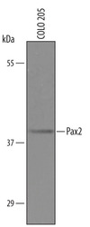
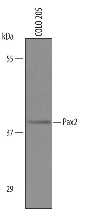
- Detection of Human Pax2 by Western Blot. Western blot shows lysates of COLO 205 human colorectal adenocarcinoma cell line. PVDF Membrane was probed with 1 µg/mL of Goat Anti-Human Pax2 Antigen Affinity-purified Polyclonal Antibody (Catalog # AF3364) followed by HRP-conjugated Anti-Goat IgG Secondary Antibody (Catalog # HAF019). A specific band was detected for Pax2 at approximately 40 kDa (as indicated). This experiment was conducted under reducing conditions and using Immunoblot Buffer Group 8.
Supportive validation
- Submitted by
- R&D Systems (provider)
- Main image
- Experimental details
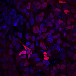
- Pax2 in BG01V Human Embyonic Stem Cells. Pax2 was detected in immersion fixed BG01V human embryonic stem cells differentiated into the early otic lineage using Goat Anti-Human Pax2 Antigen Affinity-purified Polyclonal Antibody (Catalog # AF3364) at 10 µg/mL for 3 hours at room temperature. Cells were stained using the NorthernLights™ 557-conjugated Anti-Goat IgG Secondary Antibody (red; Catalog # NL001) and counterstained with DAPI (blue). Specific staining was localized to nuclei. View our protocol for Fluorescent ICC Staining of Stem Cells on Coverslips.
