Explore
 Validate
 Learn
Learn Western blot
 ELISA
ELISAAntibody data
- Antibody Data
- Antigen structure
- References [2]
- Comments [0]
- Validations
- Western blot [1]
Submit
Validation data
Reference
Comment
Report error
- Product number
- A05032-1 - Provider product page

- Provider
- Boster Biological Technology
- Product name
- Anti-OAS3 Antibody Picoband™
- Antibody type
- Polyclonal
- Description
- Rabbit IgG polyclonal antibody for OAS3 detection. Tested with WB, ICC/IF, FCM, Direct ELISA in Human.
- Reactivity
- Human
- Host
- Rabbit
- Vial size
- 100μg/vial
- Concentration
- Add 0.2ml of distilled water will yield a concentration of 500ug/ml.
- Storage
- At -20°C for one year. After reconstitution, at 4°C for one month. It can also be aliquoted and stored frozen at -20°C for a longer time. Avoid repeated freezing and thawing.
- Handling
- Add 0.2ml of distilled water will yield a concentration of 500ug/ml.
Submitted references OAS1, OAS2, and OAS3 Contribute to Epidermal Keratinocyte Proliferation by Regulating Cell Cycle and Augmenting IFN-1‒Induced Jak1‒Signal Transducer and Activator of Transcription 1 Phosphorylation in Psoriasis.
Preparation and characterization of latex films photo-immobilized with IFN-α.
Huang YZ, Zheng YX, Zhou Y, Xu F, Cui YZ, Chen XY, Wang ZY, Yan BX, Zheng M, Man XY
The Journal of investigative dermatology 2022 Oct;142(10):2635-2645.e9
The Journal of investigative dermatology 2022 Oct;142(10):2635-2645.e9
Preparation and characterization of latex films photo-immobilized with IFN-α.
Wu L, Hu K, Zhang L, Chen W, Chen X, You R, Yin L, Guan YQ
Colloids and surfaces. B, Biointerfaces 2016 Sep 1;145:104-113
Colloids and surfaces. B, Biointerfaces 2016 Sep 1;145:104-113
No comments: Submit comment
Supportive validation
- Submitted by
- Boster Biological Technology (provider)
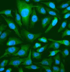
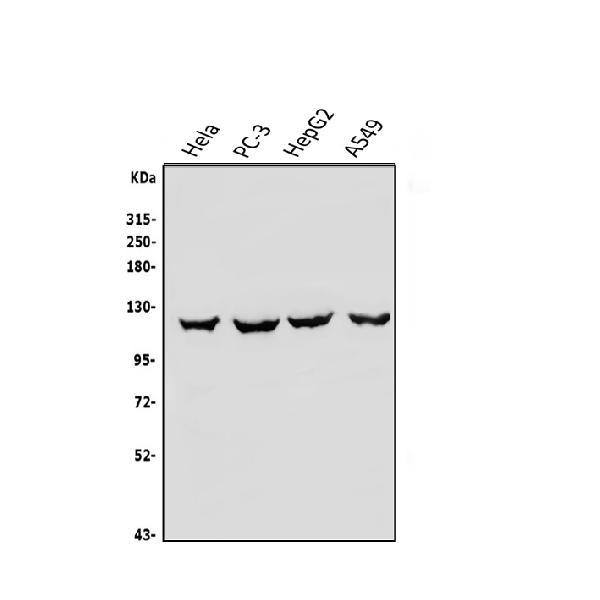
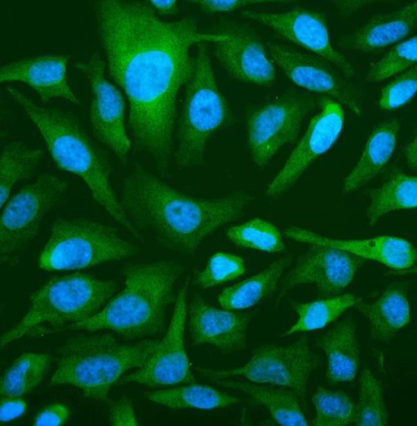
- Main image

- Experimental details
- Western blot analysis of OAS3 using anti-OAS3 antibody (A05032-1). Electrophoresis was performed on a 5-20% SDS-PAGE gel at 70V (Stacking gel) / 90V (Resolving gel) for 2-3 hours. The sample well of each lane was loaded with 50ug of sample under reducing conditions. Lane 1: human Hela whole cell lysates, Lane 2: human PC-3 whole cell lysates, Lane 3: human HepG2 whole cell lysates, Lane 4: human A549 whole cell lysates. After Electrophoresis, proteins were transferred to a Nitrocellulose membrane at 150mA for 50-90 minutes. Blocked the membrane with 5% Non-fat Milk/ TBS for 1.5 hour at RT. The membrane was incubated with rabbit anti-OAS3 antigen affinity purified polyclonal antibody (Catalog # A05032-1) at 0.25 μg/mL overnight at 4°C, then washed with TBS-0.1%Tween 3 times with 5 minutes each and probed with a goat anti-rabbit IgG-HRP secondary antibody at a dilution of 1:5000 for 1.5 hour at RT. The signal is developed using an Enhanced Chemiluminescent detection (ECL) kit (Catalog # EK1002) with Tanon 5200 system. A specific band was detected for OAS3 at approximately 123KD. The expected band size for OAS3 is at 123KD.
- Additional image