Explore
 Validate
 Learn
Learn Western blot
 Immunocytochemistry
Antibody data
- Antibody Data
- Antigen structure
- References [1]
- Comments [0]
- Validations
- Immunocytochemistry [1]
- Other assay [2]
Submit
Validation data
Reference
Comment
Report error
- Product number
- 703032 - Provider product page

- Provider
- Invitrogen Antibodies
- Product name
- TAZ Recombinant Rabbit Monoclonal Antibody (7H33L24)
- Antibody type
- Monoclonal
- Antigen
- Other
- Description
- This antibody is predicted to react with Rat, Bovine.
- Reactivity
- Human, Mouse
- Host
- Rabbit
- Isotype
- IgG
- Antibody clone number
- 7H33L24
- Vial size
- 100 μg
- Concentration
- 0.5 mg/mL
- Storage
- Store at 4°C short term. For long term storage, store at -20°C, avoiding freeze/thaw cycles.
Submitted references miR‑125a‑5p reverses epithelial‑mesenchymal transition and restores drug sensitivity by negatively regulating TAFAZZIN signaling in breast cancer.
Li D, Chen L, Zhang X, Wang Y, Huang C, Li J, He F, He W
Molecular medicine reports 2021 Nov;24(5)
Molecular medicine reports 2021 Nov;24(5)
No comments: Submit comment
Supportive validation
- Submitted by
- Invitrogen Antibodies (provider)
- Main image
- Experimental details
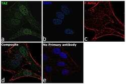
- For immunofluorescence analysis, HeLa cells were fixed and permeabilized for detection of endogenous TAZ using Anti-TAZ Recombinant Rabbit Monoclonal Antibody (Product # 703032, 1:100 dilution) and labeled with Goat anti-Rabbit IgG (H+L) Highly Cross-Adsorbed Secondary Antibody, Alexa Fluor Plus 488 conjugate (Product # A32731, 1:2000). Panel a) shows representative cells that were stained for detection and localization of TAZ (green), Panel b) is stained for nuclei (blue) using ProLong™ Diamond Antifade Mountant with DAPI (Product # P36962). Panel c) represents cytoskeletal F-actin staining using Rhodamine Phalloidin (Product # R415, 1:300). Panel d) is a composite image of Panels a, b and c clearly demonstrating Cytoplasmic and nuclear localization of TAZ. Panel e) represents control cells with no primary antibody to assess background. The images were captured at 60X magnification.

Supportive validation
- Submitted by
- Invitrogen Antibodies (provider)
- Main image
- Experimental details
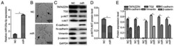
- Figure 1. miR-125a-5p reverses epithelial-mesenchymal transition in MCF7/Adr cells. (A) Transfection efficiency of mimics was determined via reverse transcription-quantitative PCR. miR-125a-5p was overexpressed after transfection of mimics in MCF-7/Adr cells. (B) Cell morphological alterations after miR-125a-5p transfection, as indicated by arrows. Images of cells in medium. The cells of NC groups remained elongated and dispersed with a mesenchymal morphology, and the cells of miR-125a-5p groups were closely packed and rounded with a cobblestone epithelial morphology; magnification, x200 (scale bar, 20 um). (C) Western blot analysis of TAFAZZIN, TG2, AKT, E-cadherin, N-cadherin, vimentin and beta-catenin in MCF7/Adr cells. (D and E) Semi-quantification of protein expression results from panel (C). GAPDH served as an internal control. Data are presented as the mean +- SD. Similar results were obtained from three independent experiments. *P

- Submitted by
- Invitrogen Antibodies (provider)
- Main image
- Experimental details
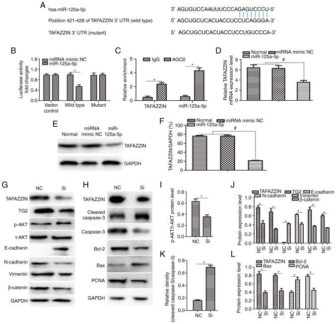
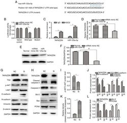
- Figure 3. TAFAZZIN is a target of miR-125a-5p in MCF-7/Adr cells. (A) Binding site of miR-125a-5p and TAFAZZIN 3'-UTR according to online bioinformatics analysis. (B) Luciferase activity was decreased for wild-type-TAFAZZIN, but not for the mutant. (C) RNA immunoprecipitation assay indicated that miR-125a-5p and TAFAZZIN were conjunct in MCF-7/Adr cell lines. (D) TAFAZZIN mRNA expression in miR-125a-5p and control groups as detected via reverse transcription-quantitative PCR. (E) Western blot analysis of TAFAZZIN protein expression in miR-125a-5p and control groups. (F) Semi-quantification of TAFAZZIN protein expression in panel (E). MCF-7/Adr cells were treated with siRNA-TAFAZZIN, and the expression levels of (G) TAFAZZIN, TG2, p-AKT, t-AKT, E-cadherin, N-cadherin, vimentin, beta-catenin, (H) cleaved caspase-3, caspase-3, pro-apoptotic protein (Bax) and PCNA were detected via western blotting. Knockdown of TAFAZZIN expression could significantly reduce TAFAZZIN, TG2, p-AKT, N-cadherin, vimentin, beta-catenin, Bcl-2 and PCNA protein expression and increase those of E-cadherin and Bax, as well as activate caspase-3. (I-L) Semi-quantification of protein levels in panels (G and H). GAPDH was used as an internal reference. Data are shown as the mean +- SD. Similar results were obtained from three independent experiments. *P