Explore
 Validate
 Learn
Learn Western blot
 Immunocytochemistry
Antibody data
- Antibody Data
- Antigen structure
- References [3]
- Comments [0]
- Validations
- Immunocytochemistry [1]
Submit
Validation data
Reference
Comment
Report error
- Product number
- PA1-936 - Provider product page

- Provider
- Invitrogen Antibodies
- Product name
- Anti-Myosin 7a
- Antibody type
- Polyclonal
- Antigen
- Synthetic Peptide: S(16) G Q E F D V P I G A V V K L C(31)
- Host
- Rabbit
Submitted references A missense mutation in the previously undescribed gene Tmhs underlies deafness in hurry-scurry (hscy) mice.
Actin-based motor properties of native myosin VIIa.
Myosin VIIa, the product of the Usher 1B syndrome gene, is concentrated in the connecting cilia of photoreceptor cells.
Longo-Guess CM, Gagnon LH, Cook SA, Wu J, Zheng QY, Johnson KR
Proceedings of the National Academy of Sciences of the United States of America 2005 May 31;102(22):7894-9
Proceedings of the National Academy of Sciences of the United States of America 2005 May 31;102(22):7894-9
Actin-based motor properties of native myosin VIIa.
Udovichenko IP, Gibbs D, Williams DS
Journal of cell science 2002 Jan 15;115(Pt 2):445-50
Journal of cell science 2002 Jan 15;115(Pt 2):445-50
Myosin VIIa, the product of the Usher 1B syndrome gene, is concentrated in the connecting cilia of photoreceptor cells.
Liu X, Vansant G, Udovichenko IP, Wolfrum U, Williams DS
Cell motility and the cytoskeleton 1997;37(3):240-52
Cell motility and the cytoskeleton 1997;37(3):240-52
No comments: Submit comment
Supportive validation
- Submitted by
- Invitrogen Antibodies (provider)
- Main image
- Experimental details
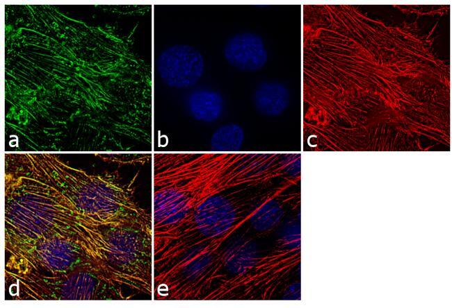
- Immunofluorescence analysis of MYO7A was performed using 70% confluent log phase L6 cells. The cells were fixed with 4% paraformaldehyde for 10 minutes, permeabilized with 0.1% Triton™ X-100 for 10 minutes, and blocked with 1% BSA for 1 hour at room temperature. The cells were labeled with MYO7A Rabbit Polyclonal Antibody (Product # PA1-936) at 2 µg/mL in 0.1% BSA and incubated for 3 hours at room temperature and then labeled with Goat anti-Rabbit IgG (Heavy Chain) Superclonal™ Secondary Antibody, Alexa Fluor® 488 conjugate (Product # A27034) at a dilution of 1:2000 for 45 minutes at room temperature (Panel a: green). Nuclei (Panel b: blue) were stained with SlowFade® Gold Antifade Mountant with DAPI (Product # S36938). F-actin (Panel c: red) was stained with Alexa Fluor® 555 Rhodamine Phalloidin (Product # R415, 1:300). Panel d represents the merged image showing cytoskeletal localization. Panel e shows the control without primary antibody. The images were captured at 60X magnification.