Explore
 Validate
 Learn
LearnGTX24864
antibody from GeneTex
Targeting: MAPT
DDPAC, FLJ31424, FTDP-17, MAPTL, MGC138549, MSTD, MTBT1, MTBT2, PPND, PPP1R103, tau
 Western blot
 Flow cytometry
Antibody data
- Antibody Data
- Antigen structure
- References [3]
- Comments [0]
- Validations
- Western blot [1]
- Immunocytochemistry [1]
- Immunohistochemistry [1]
Submit
Validation data
Reference
Comment
Report error
- Product number
- GTX24864 - Provider product page

- Provider
- GeneTex
- Proper citation
- GeneTex Cat#GTX24864, RRID:AB_373935
- Product name
- Tau (phospho Ser199/Ser202) antibody
- Antibody type
- Polyclonal
- Reactivity
- Human, Mouse, Rat
- Host
- Rabbit
Submitted references Activation of G-protein coupled estrogen receptor 1 improves early-onset cognitive impairment via PI3K/Akt pathway in rats with traumatic brain injury.
Overactivation of NR2B-containing NMDA receptors through entorhinal-hippocampal connection initiates accumulation of hyperphosphorylated tau in rat hippocampus after transient middle cerebral artery occlusion.
The role of heat shock protein 70 in the protective effect of YC-1 on β-amyloid-induced toxicity in differentiated PC12 cells.
Wang ZF, Pan ZY, Xu CS, Li ZQ
Biochemical and biophysical research communications 2017 Jan 22;482(4):948-953
Biochemical and biophysical research communications 2017 Jan 22;482(4):948-953
Overactivation of NR2B-containing NMDA receptors through entorhinal-hippocampal connection initiates accumulation of hyperphosphorylated tau in rat hippocampus after transient middle cerebral artery occlusion.
Xu CS, Liu AC, Chen J, Pan ZY, Wan Q, Li ZQ, Wang ZF
Journal of neurochemistry 2015 Aug;134(3):566-77
Journal of neurochemistry 2015 Aug;134(3):566-77
The role of heat shock protein 70 in the protective effect of YC-1 on β-amyloid-induced toxicity in differentiated PC12 cells.
Tsai YC, Lee YM, Lam KK, Lin JF, Wang JJ, Yen MH, Cheng PY
PloS one 2013;8(7):e69320
PloS one 2013;8(7):e69320
No comments: Submit comment
Supportive validation
- Submitted by
- GeneTex (provider)
- Main image
- Experimental details
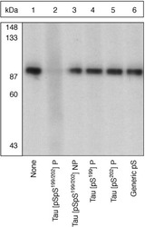
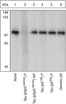
- Extracts of from African green monkey kidney (CV-1) cells were resolved by SDS-PAGE on a 10% polyacrylamide gel and transferred to nitrocellulose. Membranes were blocked with a 5% BSA-TBST buffer overnight at 4¢XC, then incubated with Tau [pSpS199/202] antibody for two hours at room temperature in a 3% BSA-TBST buffer, following prior incubation with: no peptide (1), the phosphopeptide immunogen (2), the non-phosphorylated peptide corresponding to the immunogen (3), the tau phosphopeptide corresponding to [pS199] (4), the tau phosphopeptide corresponding to [pS202] (5), or, a generic phosphoserine-containing peptide (6). After washing, membranes were incubated with goat F(ab)2 anti-rabbit IgG alkaline phosphatase conjugate and bands were detected.The data show that only the phosphopeptide corresponding to Tau [pSpS199/202] completely blocks the antibody signal, demonstrating the specificity of the antibody (GTX24864).
Supportive validation
- Submitted by
- GeneTex (provider)
- Main image
- Experimental details
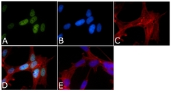
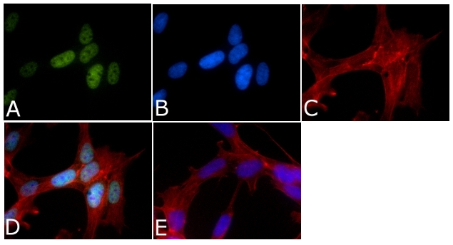
- ICC/IF analysis of SHSY5Y cells using GTX24864 Tau (phospho Ser199/Ser202) antibody. A (Green): Tau (phospho Ser199/Ser202)B (Blue): NucleiC (Red): F-actin D: Merged imageE: Without primary antibodyDilution: 1?g/mL
Supportive validation
- Submitted by
- GeneTex (provider)
- Main image
- Experimental details
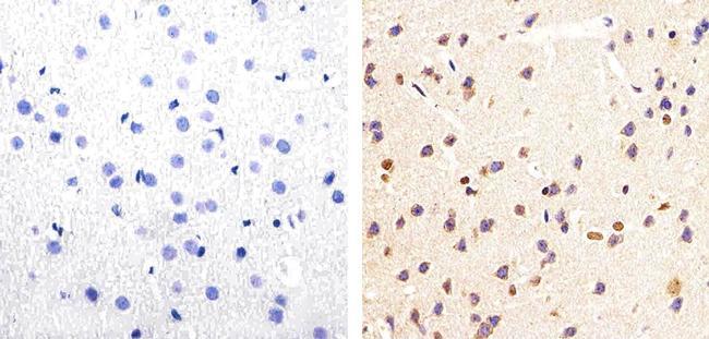
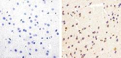
- IHC-P analysis of mouse brain tissue using GTX24864 Tau (phospho Ser199/Ser202) antibody. Left : without primary antibody Right : Tau (phospho Ser199/Ser202) antibody. Antigen retrieval : heat-induced antigen retrieval with 10mM sodium citrate (pH 6.0) for 15 min.