 Explore
 Validate
 Learn
Learn701056
antibody from Invitrogen Antibodies
Targeting: MAPT
DDPAC, FLJ31424, FTDP-17, MAPTL, MGC138549, MSTD, MTBT1, MTBT2, PPND, PPP1R103, tau
 Western blot
Antibody data
- Antibody Data
- Antigen structure
- References [3]
- Comments [0]
- Validations
- Western blot [3]
- Immunocytochemistry [1]
- Immunohistochemistry [3]
- Other assay [3]
Submit
Validation data
Reference
Comment
Report error
- Product number
- 701056 - Provider product page

- Provider
- Invitrogen Antibodies
- Product name
- Phospho-Tau (Thr231) Recombinant Rabbit Monoclonal Antibody (1H6L6)
- Antibody type
- Monoclonal
- Antigen
- Synthetic peptide
- Description
- Intact IgG appears on a non-reducing gel as ~150 kDa band and upon reduction generating a ~25 kDa light chain band and a ~50 kDa heavy chain.
- Antibody clone number
- 1H6L6
- Concentration
- 0.5 mg/mL
Submitted references Ageing and amyloidosis underlie the molecular and pathological alterations of tau in a mouse model of familial Alzheimer's disease.
Early Evidence of Low Bone Density and Decreased Serotonergic Synthesis in the Dorsal Raphe of a Tauopathy Model of Alzheimer's Disease.
Effects of antibodies to phosphorylated and non-phosphorylated tau on in vitro tau phosphorylation at Serine-199: Preliminary report.
Metaxas A, Thygesen C, Kempf SJ, Anzalone M, Vaitheeswaran R, Petersen S, Landau AM, Audrain H, Teeling JL, Darvesh S, Brooks DJ, Larsen MR, Finsen B
Scientific reports 2019 Oct 31;9(1):15758
Scientific reports 2019 Oct 31;9(1):15758
Early Evidence of Low Bone Density and Decreased Serotonergic Synthesis in the Dorsal Raphe of a Tauopathy Model of Alzheimer's Disease.
Dengler-Crish CM, Smith MA, Wilson GN
Journal of Alzheimer's disease : JAD 2017;55(4):1605-1619
Journal of Alzheimer's disease : JAD 2017;55(4):1605-1619
Effects of antibodies to phosphorylated and non-phosphorylated tau on in vitro tau phosphorylation at Serine-199: Preliminary report.
Loeffler DA, Smith LM, Klaver AC, Martić S
Experimental gerontology 2015 Jul;67:15-8
Experimental gerontology 2015 Jul;67:15-8
No comments: Submit comment
Supportive validation
- Submitted by
- Invitrogen Antibodies (provider)
- Main image
- Experimental details
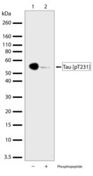
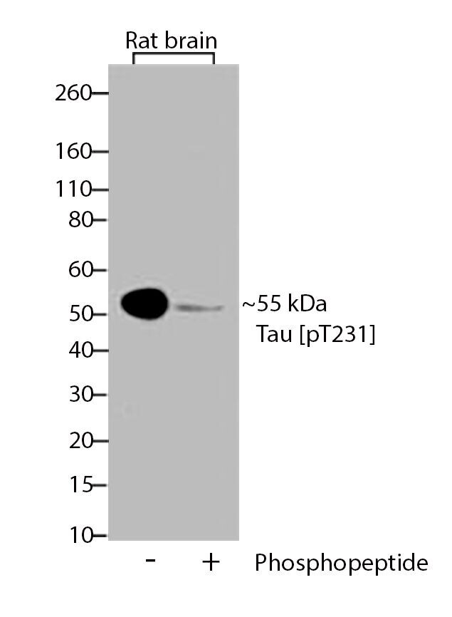
- Western blot analysis of Phospho-Tau pThr231 in total rat brain lysate using a Phospho-Tau pThr231 recombinant rabbit monoclonal antibody (Product # 701056) at a dilution of 1 µg/mL. To confirm specificity, competition was performed by preincubation with the phosphopeptide to inhibit antibody binding (lane 2). Samples were detected using chemiluminescence (ECL). Results show a band at ~56kDa.
- Submitted by
- Invitrogen Antibodies (provider)
- Main image

- Experimental details
- Western blot analysis of Phospho-Tau pThr231 in total rat brain lysate using a Phospho-Tau pThr231 recombinant rabbit monoclonal antibody (Product # 701056) at a dilution of 1 µg/mL. To confirm specificity, competition was performed by preincubation with the phosphopeptide to inhibit antibody binding (lane 2). Samples were detected using chemiluminescence (ECL). Results show a band at ~56kDa.

- Submitted by
- Invitrogen Antibodies (provider)
- Main image

- Experimental details
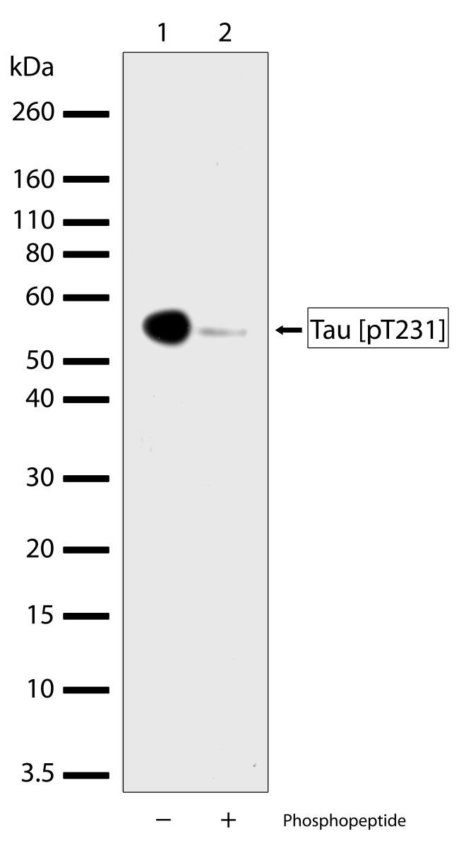
- Western blot analysis of Tau (pT231) was performed by loading 30 µg of rat brain lysate (lane 1) using Novex®NuPAGE®4-12% Bis-Tris gel (Product # NP0321BOX), XCell SureLock Electrophoresis System (Product # EI0002), Novex® Sharp Pre-Stained Protein Standard (Product # LC5800), and iBlot® Dry Blotting System (Product # IB21001). Proteins were transferred to a nitrocellulose membrane and blocked with 5% skim milk for 1 hour at room temperature. Tau (pT231) was detected at ~55 kDa using Tau (pT231) Recombinant Rabbit Monoclonal Antibody (Product # 701056) at a 1:1000 dilution in 2.5% skim milk at 4°C overnight on a rocking platform. To confirm specificity, competition was performed with the phosphopeptide (10 µg/mL) (lane 2). Detection was performed using an HRP-conjugated Goat anti-Rabbit secondary antibody (Product # G-21234) at a 1:5000 dilution and chemiluminescent detection was performed using Pierce™ ECL Western blotting Substrate (Product # 32106).
Supportive validation
- Submitted by
- Invitrogen Antibodies (provider)
- Main image

- Experimental details
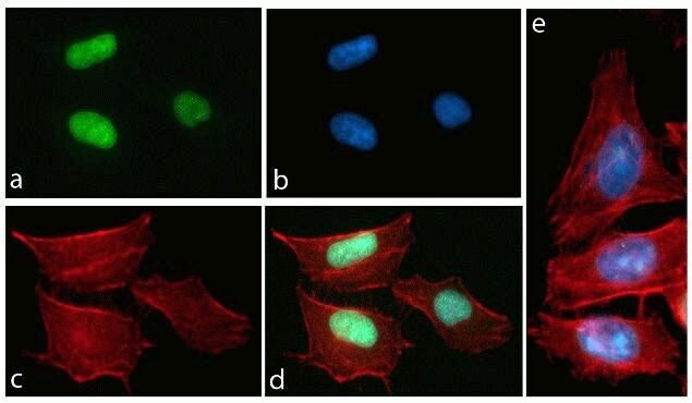
- Immunofluorescent analysis of Tau (pT231) was performed on 70% confluent log phase HeLa cells. The cells were fixed with 4% paraformaldehyde for 15 minutes, permeabilized with 0. 25% Triton X-100 for 10 minutes, and blocked with 5% BSA for 1 hour at room temperature. The cells were labeled with Tau (pT231) Recombinant Rabbit Monoclonal Antibody (Product # 701056) at a dilution of 1:500 in 1% BSA and incubated for 3 hours at room temperature and then labeled with Alexa Fluor® 488 Goat anti-Rabbit IgG secondary antibody (Product # A-11008) at a dilution of 1:400 for 30 minutes at room temperature (Panel a: green). Nuclei (Panel b: blue) were stained with SlowFade® Gold Antifade Mountant with DAPI (Product # S36938). F-actin (Panel c: red) was stained with Alexa Fluor® 594 phalloidin (Product # A12381). Panel d is a merged image showing nuclear localization and panel e shows competition with the phospho-Tau (pT231) peptide.
Supportive validation
- Submitted by
- Invitrogen Antibodies (provider)
- Main image

- Experimental details
- Immunohistochemistry analysis of Phospho-Tau pThr231 showing staining in the cytoplasm of paraffin-embedded human brain tissue (right) compared to a negative control without primary antibody (left). To expose target proteins, antigen retrieval was performed using 10 mM sodium citrate (pH 6.0), microwaved for 8-15 min. Following antigen retrieval, tissues were blocked in 3% H2O2-methanol for 15 min at room temperature, washed with ddH2O and PBS, and then probed with Phospho-Tau pThr231 (1H6L8) Monoclonal antibody (Product # 701056) diluted in 3% BSA-PBS at a dilution of 1:200 overnight at 4°C in a humidified chamber. Tissues were washed extensively in PBST and detection was performed using a HRP-conjugated secondary antibody followed by colorimetric detection using a DAB kit. Tissues were counterstained with hematoxylin and dehydrated with ethanol and xylene to prep for mounting.
- Submitted by
- Invitrogen Antibodies (provider)
- Main image

- Experimental details
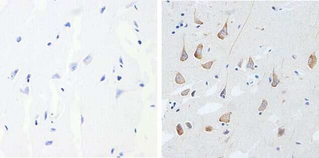
- Immunohistochemistry analysis of Phospho-Tau pThr231 showing staining in the cytoplasm of paraffin-embedded human glioma (right) compared to a negative control without primary antibody (left). To expose target proteins, antigen retrieval was performed using 10 mM sodium citrate (pH 6.0), microwaved for 8-15 min. Following antigen retrieval, tissues were blocked in 3% H2O2-methanol for 15 min at room temperature, washed with ddH2O and PBS, and then probed with a Phospho-Tau pThr231 (1H6L8) Monoclonal antibody (Product # 701056) diluted in 3% BSA-PBS at a dilution of 1:200 overnight at 4°C in a humidified chamber. Tissues were washed extensively in PBST and detection was performed using a HRP-conjugated secondary antibody followed by colorimetric detection using a DAB kit. Tissues were counterstained with hematoxylin and dehydrated with ethanol and xylene to prep for mounting.
- Submitted by
- Invitrogen Antibodies (provider)
- Main image

- Experimental details
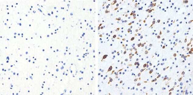
- Immunohistochemistry analysis of Phospho-Tau pThr231 showing staining in the cytoplasm of paraffin-embedded rat brain tissue (right) compared to a negative control without primary antibody (left). To expose target proteins, antigen retrieval was performed using 10 mM sodium citrate (pH 6.0), microwaved for 8-15 min. Following antigen retrieval, tissues were blocked in 3% H2O2-methanol for 15 min at room temperature, washed with ddH2O and PBS, and then probed with a Phospho-Tau pThr231 (1H6L8) Monoclonal antibody (Product # 701056) diluted in 3% BSA-PBS at a dilution of 1:200 overnight at 4°C in a humidified chamber. Tissues were washed extensively in PBST and detection was performed using a HRP-conjugated secondary antibody followed by colorimetric detection using a DAB kit. Tissues were counterstained with hematoxylin and dehydrated with ethanol and xylene to prep for mounting.

Supportive validation
- Submitted by
- Invitrogen Antibodies (provider)
- Main image
- Experimental details
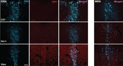
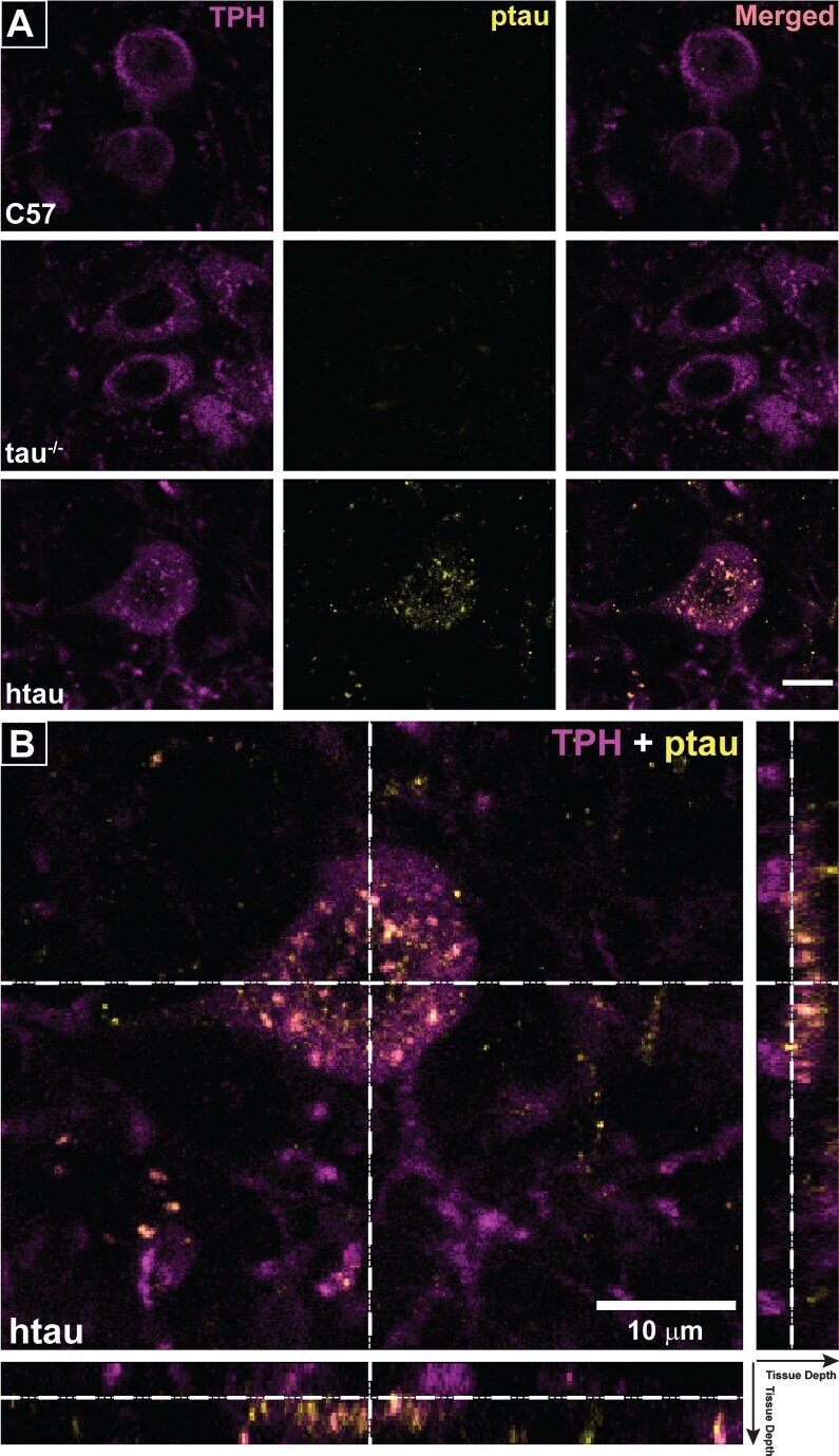
- Fig.4 Phosphorylated tau (ptau) accumulation in trytophan hydroxylase (TPH)-positive cells of the DRN is evident in 4-month-old htau mice. Photomicrographs of immunofluorescent-stained DRN sections are shown in the first three columns for C57, tau -/- , and htau mice. All mice exhibit distinctly-labeled TPH cells; however, ptau-231 label is non-existent in C57 and tau -/- DRN. ptau-231 immunofluorescence is obvious in DRN sections of 4-month-old htau mice with substantial co-label of this marker in TPH-positive cells. Fourth column: Photomicrographs of merged channels of TPH and ptau for MRN from each respective animal. Note that ptau-231 fluorescence in htau MRN is weak compared to DRN. Scale bar = 100 mum.

- Submitted by
- Invitrogen Antibodies (provider)
- Main image
- Experimental details
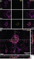
- Fig.5 Co-localization of ptau-231 in tryptophan hydroxylase (TPH)-positive cells of 4-month-old htau DRN. A) Confocal images of single planes through rostral DRN at the level of the trochlear nuclei for C57, tau -/- , and htau mice. Phosphorylated tau (ptau) fluorescence is co-localized to TPH-positive cells in the htau section only. Scale bar = 10 mum. B) 3-D reconstruction of a TPH-positive cell in the htau DRN. Orthogonal views of the stack depict fluorescence label across tissue depth. Crosshairs (dashed lines) indicate the plane of section for the X, Y, and Z dimensions. The TPH-positive cell shows prominent ptau label in the cytoplasm of the soma with sparser puncta in TPH-positive processes.Scale bar = 10 mum.
- Submitted by
- Invitrogen Antibodies (provider)
- Main image
- Experimental details
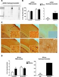
- Figure 6 Validation of pS404, pT231 and 3R tau. ( A) Immunoblotting of sarkosyl-insoluble tau with rabbit primary antibody against phospho-Ser404 (1:200; OAAF07796, Aviva Systems Biology). The entire membrane is shown. Hyperphosphorylation at the S404 residue was exclusively observed in 24-month-old APP swe /PS1 DeltaE9 mice (TG 24 M, lane 8). Lanes are labelled as follows: Marker: 1, 14; AD: 3; non-AD: 5; WT 24 months: 6; TG 24 months: 8; WT 3 months: 10; TG 3 months: 12; Empty: 2, 4, 7, 9, 11, 13. (B) ELISA of pT231 tau. Soluble pT231 tau was present in the neocortex of both WT and TG mice. Phosphorylation at T231 was only observed in the sarkosyl-insoluble fraction from 24-month-old APP swe /PS1 DeltaE9 mice. Results are expressed as arbitrary units (U), normalized to total protein concentration. (C) Immunohistochemistry of pT231 tau in coronal, 50 um-thick brain sections from 3- and 18-month-old WT and TG mice. Black arrows point to CA1 pyramidal neurons, which were more strongly immunolabelled in 18 vs. 3-month-old animals, irrespective of genotype. Scale bar: 200 um. The inserts show higher magnifications of layer V neurons in the temporal cortex, with black arrowheads pointing to tangle-like structures in aged APP swe /PS1 DeltaE9 mice. Note reduced dendritic staining in 18 vs. 3-month-old animals. Scale bar: 10 um. White arrows point to puncta of pT231 immunoreactivity within a plaque-like structure, observed exclusively in the neocortex of TG mice. Scale b
