Explore
 Validate
 Learn
Learn710298
antibody from Invitrogen Antibodies
Targeting: MAPT
DDPAC, FLJ31424, FTDP-17, MAPTL, MGC138549, MSTD, MTBT1, MTBT2, PPND, PPP1R103, tau
 Western blot
Antibody data
- Antibody Data
- Antigen structure
- References [1]
- Comments [0]
- Validations
- Western blot [2]
- Immunocytochemistry [1]
- Immunohistochemistry [1]
- Other assay [1]
Submit
Validation data
Reference
Comment
Report error
- Product number
- 710298 - Provider product page

- Provider
- Invitrogen Antibodies
- Product name
- Phospho-Tau (Ser396) Recombinant Polyclonal Antibody (5HCLC)
- Antibody type
- Polyclonal
- Antigen
- Synthetic peptide
- Description
- This antibody is predicted to react with mouse, rabbit and non-human primate based on sequence homology.
- Antibody clone number
- 5HCLC
- Concentration
- 0.5 mg/mL
Submitted references Dihydrolipoamide dehydrogenase suppression induces human tau phosphorylation by increasing whole body glucose levels in a C. elegans model of Alzheimer's Disease.
Ahmad W
Experimental brain research 2018 Nov;236(11):2857-2866
Experimental brain research 2018 Nov;236(11):2857-2866
No comments: Submit comment
Supportive validation
- Submitted by
- Invitrogen Antibodies (provider)
- Main image
- Experimental details
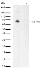
- Western blot analysis of Phospho-Tau pSer396 in rat brain lysate using a Phospho-Tau pSer396 Recombinant Rabbit Polyclonal Antibody (Product # 710298) at a dilution of 2 µg/mL. To confirm specificity, competition was performed by preincubation with the phosphopeptide to inhibit antibody binding (lane 2). Samples were detected using chemiluminescence (ECL). Results show a band at ~76kDa.

- Submitted by
- Invitrogen Antibodies (provider)
- Main image
- Experimental details
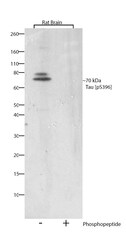
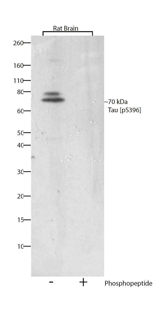
- Western blot analysis of Tau (pS396) was performed by loading 30 µg of rat brain lysate (lane 1) using Novex®NuPAGE® 4-12% Bis-Tris gel (Product # NP0321BOX), XCell SureLock Electrophoresis System (Product # EI0002), Novex® Sharp Pre-Stained Protein Standard (Product # LC5800), and iBlot® Dry Blotting System (Product # IB21001). Proteins were transferred to a nitrocellulose membrane and blocked with 5% skim milk for 1 hour at room temperature. Tau (pS396) was detected at ~70 kDa using Tau (pS396) Recombinant Rabbit Polyclonal Antibody (Product # 710298) at a 1:1000 dilution in 2.5% skim milk at 4°C overnight on a rocking platform. To confirm specificity, competition was performed with the phosphopeptide (10 µg/mL) (lane 2). Detection was performed using an HRP-conjugated Goat anti-Rabbit secondary antibody (Product # G-21234) at a 1:5000 dilution and chemiluminescent detection was performed using Pierce™ ECL Western blotting Substrate (Product # 32106).
Supportive validation
- Submitted by
- Invitrogen Antibodies (provider)
- Main image
- Experimental details
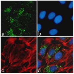
- Immunofluorescent analysis of Tau (pS396) was performed on 70% confluent log phase HeLa cells. The cells were fixed with 4% paraformaldehyde for 15 minutes, permeabilized with 0. 25% Triton X-100 for 10 minutes, and blocked with 5% BSA for 1 hour at room temperature. The cells were labeled with Tau (pS396) Recombinant Rabbit Polyclonal Antibody (Product # 710298) at a dilution of 1:500 in 1% BSA and incubated for 3 hours at room temperature and then labeled with Alexa Fluor® 488 Goat anti-Rabbit IgG secondary antibody (Product # A-11008) at a dilution of 1:400 for 30 minutes at room temperature (Panel a: green). Nuclei (Panel b: blue) were stained with SlowFade® Gold Antifade Mountant with DAPI (Product # S36938). F-actin (Panel c: red) was stained with Alexa Fluor® 594 phalloidin (Product # A12381). Panel d is a merged image showing cytoplasmic localization. The images were captured using a Nikon microscope at 20X magnification.
Supportive validation
- Submitted by
- Invitrogen Antibodies (provider)
- Main image
- Experimental details
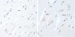
- Immunohistochemistry analysis of Phospho-Tau pSer396 showing staining in the cytoplasm of paraffin-embedded human brain tissue (right) compared to a negative control without primary antibody (left). To expose target proteins, antigen retrieval was performed using 10 mM sodium citrate (pH 6.0), microwaved for 8-15 min. Following antigen retrieval, tissues were blocked in 3% H2O2-methanol for 15 min at room temperature, washed with ddH2O and PBS, and then probed with Phospho-Tau pSer396 Antibody (5HCLC) Recombinant Rabbit Polyclonal Antibody (Product # 710298) diluted in 3% BSA-PBS at a dilution of 1:20 overnight at 4°C in a humidified chamber. Tissues were washed extensively in PBST and detection was performed using a HRP-conjugated secondary antibody followed by colorimetric detection using a DAB kit. Tissues were counterstained with hematoxylin and dehydrated with ethanol and xylene to prep for mounting.
Supportive validation
- Submitted by
- Invitrogen Antibodies (provider)
- Main image

- Experimental details
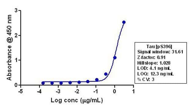
- Indirect ELISA analysis of Phospho-Tau pSer396 in rat brain lysate coated onto the plate using a Phospho-Tau pSer396 Recombinant Rabbit Polyclonal Antibody (Product # 710298) at various dilutions. A non-linear regression analysis was performed (4 PL) and LOD and LOQ for the antibody were determined.