 Explore
 Validate
 Learn
LearnPA5-27287
antibody from Invitrogen Antibodies
Targeting: MAPT
DDPAC, FLJ31424, FTDP-17, MAPTL, MGC138549, MSTD, MTBT1, MTBT2, PPND, PPP1R103, tau
 Western blot
Antibody data
- Antibody Data
- Antigen structure
- References [3]
- Comments [0]
- Validations
- Western blot [5]
- Immunohistochemistry [3]
- Other assay [1]
Submit
Validation data
Reference
Comment
Report error
- Product number
- PA5-27287 - Provider product page

- Provider
- Invitrogen Antibodies
- Product name
- Tau Polyclonal Antibody
- Antibody type
- Polyclonal
- Antigen
- Recombinant protein fragment
- Description
- Recommended positive controls: SK-N-SH, IMR32, SK-N-AS, mouse brain. Predicted reactivity: Chimpanzee (99%). Store product as a concentrated solution. Centrifuge briefly prior to opening the vial.
- Reactivity
- Human, Mouse
- Host
- Rabbit
- Isotype
- IgG
- Vial size
- 100 µL
- Concentration
- 0.34 mg/mL
- Storage
- Store at 4°C short term. For long term storage, store at -20°C, avoiding freeze/thaw cycles.
Submitted references Isolation and characterization of antibody fragment selective for human Alzheimer's disease brain-derived tau variants.
Comparison of (18)F-T807 and (18)F-THK5117 PET in a Mouse Model of Tau Pathology.
Eicosanoyl-5-hydroxytryptamide (EHT) prevents Alzheimer's disease-related cognitive and electrophysiological impairments in mice exposed to elevated concentrations of oligomeric beta-amyloid.
Venkataraman L, He P, Schulz P, Sierks MR
Neurobiology of aging 2020 Oct;94:7-14
Neurobiology of aging 2020 Oct;94:7-14
Comparison of (18)F-T807 and (18)F-THK5117 PET in a Mouse Model of Tau Pathology.
Brendel M, Yousefi BH, Blume T, Herz M, Focke C, Deussing M, Peters F, Lindner S, von Ungern-Sternberg B, Drzezga A, Bartenstein P, Haass C, Okamura N, Herms J, Yakushev I, Rominger A
Frontiers in aging neuroscience 2018;10:174
Frontiers in aging neuroscience 2018;10:174
Eicosanoyl-5-hydroxytryptamide (EHT) prevents Alzheimer's disease-related cognitive and electrophysiological impairments in mice exposed to elevated concentrations of oligomeric beta-amyloid.
Asam K, Staniszewski A, Zhang H, Melideo SL, Mazzeo A, Voronkov M, Huber KL, Pérez E, Stock M, Stock JB, Arancio O, Nicholls RE
PloS one 2017;12(12):e0189413
PloS one 2017;12(12):e0189413
No comments: Submit comment
Supportive validation
- Submitted by
- Invitrogen Antibodies (provider)
- Main image

- Experimental details
- Western blot analysis of Tau using A) 30 µg SK-N-SH whole cell lysate (B) 30 µg IMR32 whole cell lysate and C) 30 µg SK-N-AS whole cell lysate. Samples were loaded onto a 10% SDS-PAGE gel and probed with a Tau polyclonal antibody (Product # PA5-27287) at a dilution of 1:500.

- Submitted by
- Invitrogen Antibodies (provider)
- Main image

- Experimental details
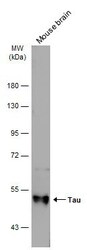
- Western blot analysis of Tau using 50 µg of mouse brain lysate. Samples were loaded onto a 10% SDS-PAGE gel and probed with a Tau polyclonal antibody (Product # PA5-27287) at a dilution of 1:3000.

- Submitted by
- Invitrogen Antibodies (provider)
- Main image

- Experimental details
- Western Blot analysis of Tau was performed by separating 30 µg of various whole cell extracts by 7.5% SDS-PAGE. Proteins were transferred to a membrane and probed with a Tau Polyclonal Antibody (Product # PA5-27287) at a dilution of 1:1000 and a HRP-conjugated anti-rabbit IgG secondary antibody.

- Submitted by
- Invitrogen Antibodies (provider)
- Main image
- Experimental details
- Western Blot analysis of Tau was performed by separating 50 µg of Mouse tissue extracts by 7.5% SDS-PAGE. Proteins were transferred to a membrane and probed with a Tau Polyclonal Antibody (Product # PA5-27287) at a dilution of 1:3000. The HRP-conjugated anti-rabbit IgG antibody was used to detect the primary antibody.

- Submitted by
- Invitrogen Antibodies (provider)
- Main image
- Experimental details
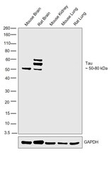
- Western blot was performed using Anti-Tau Polyclonal Antibody (Product # PA5-27287) and 40-80 kDa bands corresponding to Tau were observed across in Mouse and Rat Brain but not in Mouse Kidney or Mouse and Rat Lung. Tissue extracts (30 µg lysate) of Mouse Brain (Lane 1), Rat Brain (Lane 2), Mouse Kidney (Lane 3), Mouse Lung (Lane 4) or Rat Lung (Lane 5) were electrophoresed using NuPAGE™ 10% Bis-Tris Protein Gel (Product # NP0301BOX). Resolved proteins were then transferred onto a Nitrocellulose membrane (Product # IB23001) by iBlot® 2 Dry Blotting System (Product # IB21001). The blot was probed with the primary antibody (1:500) and detected by chemiluminescence with Goat anti-Rabbit IgG (H+L) Superclonal™ Recombinant Secondary Antibody, HRP (Product # A27036,1:4000) using the iBright FL 1000 (Product # A32752). Chemiluminescent detection was performed using Novex® ECL Chemiluminescent Substrate Reagent Kit (Product # WP20005).

Supportive validation
- Submitted by
- Invitrogen Antibodies (provider)
- Main image
- Experimental details
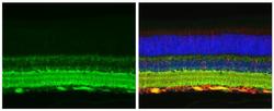
- Immunohistochemistry (Paraffin) analysis of Tau was performed in paraffin-embedded mouse eye tissue using Green: Tau Polyclonal Antibody (Product # PA5-29610) at a dilution of 1:500. Red: beta Tubulin 3/ Tuj1, a cytoskeleton marker, stained by beta Tubulin 3/ Tuj1 antibody diluted at 1:500. Blue: Fluoroshield with DAPI. Antigen Retrieval: Citrate buffer, pH 6.0, 15 min.

- Submitted by
- Invitrogen Antibodies (provider)
- Main image

- Experimental details
- Immunohistochemistry (Paraffin) analysis of Tau was performed in paraffin-embedded mouse eye tissue using Green: Tau Polyclonal Antibody (Product # PA5-29610) at a dilution of 1:500. Red: beta Tubulin 3/ Tuj1, a cytoskeleton marker, stained by beta Tubulin 3/ Tuj1 antibody diluted at 1:500. Blue: Fluoroshield with DAPI. Antigen Retrieval: Citrate buffer, pH 6.0, 15 min.

- Submitted by
- Invitrogen Antibodies (provider)
- Main image
- Experimental details
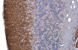
- Immunohistochemistry (Paraffin) analysis of Tau was performed in paraffin-embedded mouse cerebellum tissue using Tau Polyclonal Antibody (Product # PA5-27287) at a dilution of 1:500. Antigen Retrieval: Citrate buffer, pH 6.0, 15 min.

Supportive validation
- Submitted by
- Invitrogen Antibodies (provider)
- Main image

- Experimental details
- FIGURE 6 (A) PET-Immunohistochemistry correlation of 18 F-T807 (blue diamonds) and 18 F-THK5117 (yellow circles) SUVR with %tau/area to PA5-27287 antibody staining. (B) 3-dimensional brainstem acquisition of PA5-27287 staining and (C) zoomed image indicating automatically detected tau positive cell somata. (D) 2-dimensional whole brain overview shows tau-positive cells predominantly in the brainstem, and lower amounts in the neocortex. Data derive from N = 5 P301S mice.
