Explore
 Validate
 Learn
LearnMAB3494
antibody from R&D Systems
Targeting: MAPT
DDPAC, FLJ31424, FTDP-17, MAPTL, MGC138549, MSTD, MTBT1, MTBT2, PPND, PPP1R103, tau
 Western blot
 Immunohistochemistry
Antibody data
- Antibody Data
- Antigen structure
- References [0]
- Comments [0]
- Validations
- Immunohistochemistry [1]
Submit
Validation data
Reference
Comment
Report error
- Product number
- MAB3494 - Provider product page

- Provider
- R&D Systems
- Product name
- Tau Antibody
- Antibody type
- Monoclonal
- Description
- Protein A or G purified from hybridoma culture supernatant. Detects human Tau in direct ELISAs and Western blots.
- Host
- Mouse
- Conjugate
- Unconjugated
- Antigen sequence
P10636- Isotype
- IgG
- Antibody clone number
- 376720
- Vial size
- 100 ug
- Concentration
- LYOPH
- Storage
- Use a manual defrost freezer and avoid repeated freeze-thaw cycles. 12 months from date of receipt, -20 to -70 °C as supplied. 1 month, 2 to 8 °C under sterile conditions after reconstitution. 6 months, -20 to -70 °C under sterile conditions after reconstitution.
No comments: Submit comment
Supportive validation
- Submitted by
- R&D Systems (provider)
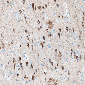
- Main image
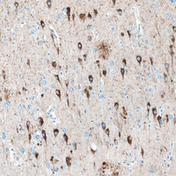
- Experimental details
- Tau in Human Brain. Tau was detected in immersion fixed paraffin-embedded sections of human Alzheimer's brain using Mouse Anti-Tau Monoclonal Antibody (Catalog # MAB3494) at 15 µg/mL overnight at 4 °C. Before incubation with the primary antibody, tissue was subjected to heat-induced epitope retrieval using Antigen Retrieval Reagent-Basic (Catalog # CTS013). Tissue was stained using the Anti-Mouse HRP-DAB Cell & Tissue Staining Kit (brown; Catalog # CTS002) and counterstained with hematoxylin (blue). Specific staining was localized to neuronal cell bodies and processes. View our protocol for Chromogenic IHC Staining of Paraffin-embedded Tissue Sections.