Explore
 Validate
 Learn
Learn14-0299-80
antibody from Invitrogen Antibodies
Targeting: ITGB1
CD29, FNRB, GPIIA, MDF2, MSK12
 Western blot
 Immunocytochemistry
 Immunoprecipitation Immunohistochemistry Flow cytometry Other assay
Antibody data
- Antibody Data
- Antigen structure
- References [10]
- Comments [0]
- Validations
- Western blot [1]
- Flow cytometry [1]
Submit
Validation data
Reference
Comment
Report error
- Product number
- 14-0299-80 - Provider product page

- Provider
- Invitrogen Antibodies
- Product name
- Anti-CD29 (Integrin beta 1) Monoclonal Antibody (TS2/16), eBioscience™
- Antibody type
- Monoclonal
- Antigen
- Other
- Description
- Description: The TS2/16 monoclonal antibody reacts with human CD29, also known as integrin beta 1, an approximately 130 kDa single-pass transmembrane glycoprotein. CD29 complexes with one of nine integrin alpha subunits to form the very late antigen (VLA) subfamily of adhesion molecules. Integrin heterodimers containing CD29 are involved in cell-cell and cell-matrix adhesion. CD29 is expressed broadly on lymphocytes and monocytes, with lower levels of expression on granulocytes. The TS2/16 antibody has been found to possess activating activity for beta 1 integrins. Applications Reported: This TS2/16 antibody has been reported for use in flow cytometric analysis, immunoprecipitation, and immunohistochemical staining. Applications Tested: This TS2/16 antibody has been tested by flow cytometric analysis of normal human peripheral blood cells. This can be used at less than or equal to 0.25 µg per test. A test is defined as the amount (µg) of antibody that will stain a cell sample in a final volume of 100 µL. Cell number should be determined empirically but can range from 10^5 to 10^8 cells/test. It is recommended that the antibody be carefully titrated for optimal performance in the assay of interest. Purity: Greater than 90%, as determined by SDS-PAGE. Aggregation: Less than 10%, as determined by HPLC. Filtration: 0.2 µm post-manufacturing filtered.
- Reactivity
- Human
- Host
- Mouse
- Isotype
- IgG
- Antibody clone number
- TS2/16
- Vial size
- 25 µg
- Concentration
- 0.5 mg/mL
- Storage
- 4° C
Submitted references Accelerated Wound Healing by Fibroblasts Differentiated from Human Embryonic Stem Cell-Derived Mesenchymal Stem Cells in a Pressure Ulcer Animal Model.
The scaffold protein p140Cap limits ERBB2-mediated breast cancer progression interfering with Rac GTPase-controlled circuitries.
Human adipose-derived stem cells partially rescue the stroke syndromes by promoting spatial learning and memory in mouse middle cerebral artery occlusion model.
Tumor-penetrating iRGD peptide inhibits metastasis.
PDGFR-β (+) perivascular cells from infantile hemangioma display the features of mesenchymal stem cells and show stronger adipogenic potential in vitro and in vivo.
Annexin A2 regulates β1 integrin internalization and intestinal epithelial cell migration.
GATA3 suppresses metastasis and modulates the tumour microenvironment by regulating microRNA-29b expression.
GMP-compliant isolation and large-scale expansion of bone marrow-derived MSC.
Sequential regulation of alpha 4 beta 1 and alpha 5 beta 1 integrin avidity by CC chemokines in monocytes: implications for transendothelial chemotaxis.
Glycoproteins of 210,000 and 130,000 m.w. on activated T cells: cell distribution and antigenic relation to components on resting cells and T cell lines.
Yoon D, Yoon D, Sim H, Hwang I, Lee JS, Chun W
Stem cells international 2018;2018:4789568
Stem cells international 2018;2018:4789568
The scaffold protein p140Cap limits ERBB2-mediated breast cancer progression interfering with Rac GTPase-controlled circuitries.
Grasso S, Chapelle J, Salemme V, Aramu S, Russo I, Vitale N, Verdun di Cantogno L, Dallaglio K, Castellano I, Amici A, Centonze G, Sharma N, Lunardi S, Cabodi S, Cavallo F, Lamolinara A, Stramucci L, Moiso E, Provero P, Albini A, Sapino A, Staaf J, Di Fiore PP, Bertalot G, Pece S, Tosoni D, Confalonieri S, Iezzi M, Di Stefano P, Turco E, Defilippi P
Nature communications 2017 Mar 16;8:14797
Nature communications 2017 Mar 16;8:14797
Human adipose-derived stem cells partially rescue the stroke syndromes by promoting spatial learning and memory in mouse middle cerebral artery occlusion model.
Zhou F, Gao S, Wang L, Sun C, Chen L, Yuan P, Zhao H, Yi Y, Qin Y, Dong Z, Cao L, Ren H, Zhu L, Li Q, Lu B, Liang A, Xu GT, Zhu H, Gao Z, Ma J, Xu J, Chen X
Stem cell research & therapy 2015 May 9;6:92
Stem cell research & therapy 2015 May 9;6:92
Tumor-penetrating iRGD peptide inhibits metastasis.
Sugahara KN, Braun GB, de Mendoza TH, Kotamraju VR, French RP, Lowy AM, Teesalu T, Ruoslahti E
Molecular cancer therapeutics 2015 Jan;14(1):120-8
Molecular cancer therapeutics 2015 Jan;14(1):120-8
PDGFR-β (+) perivascular cells from infantile hemangioma display the features of mesenchymal stem cells and show stronger adipogenic potential in vitro and in vivo.
Yuan SM, Guo Y, Zhou XJ, Shen WM, Chen HN
International journal of clinical and experimental pathology 2014;7(6):2861-70
International journal of clinical and experimental pathology 2014;7(6):2861-70
Annexin A2 regulates β1 integrin internalization and intestinal epithelial cell migration.
Rankin CR, Hilgarth RS, Leoni G, Kwon M, Den Beste KA, Parkos CA, Nusrat A
The Journal of biological chemistry 2013 May 24;288(21):15229-39
The Journal of biological chemistry 2013 May 24;288(21):15229-39
GATA3 suppresses metastasis and modulates the tumour microenvironment by regulating microRNA-29b expression.
Chou J, Lin JH, Brenot A, Kim JW, Provot S, Werb Z
Nature cell biology 2013 Feb;15(2):201-13
Nature cell biology 2013 Feb;15(2):201-13
GMP-compliant isolation and large-scale expansion of bone marrow-derived MSC.
Fekete N, Rojewski MT, Fürst D, Kreja L, Ignatius A, Dausend J, Schrezenmeier H
PloS one 2012;7(8):e43255
PloS one 2012;7(8):e43255
Sequential regulation of alpha 4 beta 1 and alpha 5 beta 1 integrin avidity by CC chemokines in monocytes: implications for transendothelial chemotaxis.
Weber C, Alon R, Moser B, Springer TA
The Journal of cell biology 1996 Aug;134(4):1063-73
The Journal of cell biology 1996 Aug;134(4):1063-73
Glycoproteins of 210,000 and 130,000 m.w. on activated T cells: cell distribution and antigenic relation to components on resting cells and T cell lines.
Hemler ME, Sanchez-Madrid F, Flotte TJ, Krensky AM, Burakoff SJ, Bhan AK, Springer TA, Strominger JL
Journal of immunology (Baltimore, Md. : 1950) 1984 Jun;132(6):3011-8
Journal of immunology (Baltimore, Md. : 1950) 1984 Jun;132(6):3011-8
No comments: Submit comment
Supportive validation
- Submitted by
- Invitrogen Antibodies (provider)
- Main image
- Experimental details
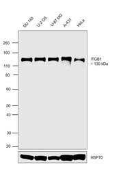
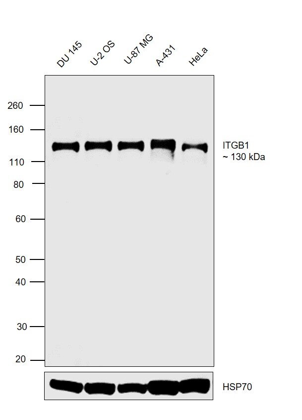
- Western blot was performed using Anti-CD29 (Integrin beta 1) Monoclonal Antibody (TS2/16), eBioscience™ (Product # 14-0299-80) and a 130 kDa band corresponding to CD29 (Integrin beta 1) was observed across the cell lines tested. Membrane enriched extracts (30 µg lysate) of DU 145 (Lane 1), U-2 OS (Lane 2), U-87 MG (Lane 3), A-431 (Lane 4) and HeLa (Lane 5) were electrophoresed using NuPAGE™ 4-12% Bis-Tris Protein Gel (Product # NP0322BOX). Resolved proteins were then transferred onto a Nitrocellulose membrane (Product # IB23001) by iBlot® 2 Dry Blotting System (Product # IB21001). The blot was probed with the primary antibody (1:1000 dilution) and detected by chemiluminescence with Goat anti-Mouse IgG (H+L) Superclonal™ Recombinant Secondary Antibody, HRP (Product # A28177,1:4000 dilution) using the iBright FL 1000 (Product # A32752). Chemiluminescent detection was performed using Novex® ECL Chemiluminescent Substrate Reagent Kit (Product # WP20005).
Supportive validation
- Submitted by
- Invitrogen Antibodies (provider)
- Main image
- Experimental details
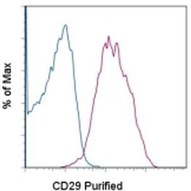
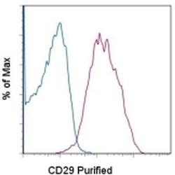
- Staining of normal human peripheral blood cells with 0.125 µg of Mouse IgG1 kappa Isotype Control Purified (Product # 14-4714-82) (blue histogram) or 0.125 µg of Anti-Human CD29 Purified (purple histogram) followed by F (ab')2 Anti-Mouse IgG PE (Product # 12-4012). Cells in the lymphocyte gate were used for analysis.