 Explore
 Validate
 Learn
Learn Western blot
Antibody data
- Antibody Data
- Antigen structure
- References [2]
- Comments [0]
- Validations
- Western blot [3]
- Immunocytochemistry [2]
- Flow cytometry [3]
Submit
Validation data
Reference
Comment
Report error
- Product number
- AF1778 - Provider product page

- Provider
- R&D Systems
- Product name
- Human/Canine Integrin beta 1/CD29 Antibody
- Antibody type
- Polyclonal
- Description
- Antigen Affinity-purified. Detects human Integrin beta 1/CD29 in direct ELISAs and Western blots. In direct ELISAs, approximately 25% cross-reactivity with recombinant mouse (rm) Integrin beta 1 is observed and less than 1% cross-reactivity with recombinant human (rh) Integrin beta 2, rhIntegrin beta 3, rhIntegrin beta 5, and rhIntegrin beta 7 is observed.
- Reactivity
- Human, Canine
- Host
- Goat
- Conjugate
- Unconjugated
- Antigen sequence
P05556- Isotype
- IgG
- Vial size
- 100 ug
- Concentration
- LYOPH
- Storage
- Use a manual defrost freezer and avoid repeated freeze-thaw cycles. 12 months from date of receipt, -20 to -70 °C as supplied. 1 month, 2 to 8 °C under sterile conditions after reconstitution. 6 months, -20 to -70 °C under sterile conditions after reconstitution.
Submitted references Bystander cells enhance NK cytotoxic efficiency by reducing search time.
In vivo biomarker expression patterns are preserved in 3D cultures of Prostate Cancer.
Zhou X, Zhao R, Schwarz K, Mangeat M, Schwarz EC, Hamed M, Bogeski I, Helms V, Rieger H, Qu B
Scientific reports 2017 Mar 13;7:44357
Scientific reports 2017 Mar 13;7:44357
In vivo biomarker expression patterns are preserved in 3D cultures of Prostate Cancer.
Windus LC, Kiss DL, Glover T, Avery VM
Experimental cell research 2012 Nov 15;318(19):2507-19
Experimental cell research 2012 Nov 15;318(19):2507-19
No comments: Submit comment
Supportive validation
- Submitted by
- R&D Systems (provider)
- Main image
- Experimental details
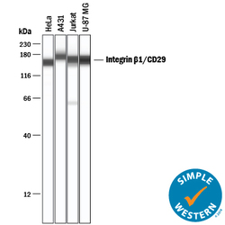
- Detection of Human Integrin beta 1/CD29 by Simple WesternTM. Simple Western lane view shows lysates of HeLa human cervical epithelial carcinoma cell line, A431 human epithelial carcinoma cell line, Jurkat human acute T cell leukemia cell line, and U-87 MG human glioblastoma/astrocytoma cell line, loaded at 0.2 mg/mL. Specific bands were detected for Integrin beta 1/CD29 at approximately 157-176 kDa (as indicated) using 10 µg/mL of Goat Anti-Human/Canine Integrin beta 1/CD29 Antigen Affinity-purified Polyclonal Antibody (Catalog # AF1778) followed by 1:50 dilution of HRP-conjugated Anti-Goat IgG Secondary Antibody (Catalog # HAF109). This experiment was conducted under reducing conditions and using the 12-230 kDa separation system.

- Submitted by
- R&D Systems (provider)
- Main image
- Experimental details
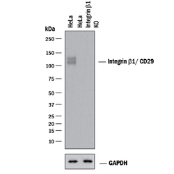
- Western Blot Shows Human Integrin beta 1/CD29 Specificity by Using Knockout Cell Line. Western blot shows lysates of HeLa human cervical epithelial carcinoma parental cell line and Integrin beta 1/CD29 knockout HeLa cell line (KO). PVDF membrane was probed with 2 µg/mL of Goat Anti-Human/Canine Integrin beta 1/CD29 Antigen Affinity-purified Polyclonal Antibody (Catalog # AF1778) followed by HRP-conjugated Anti-Goat IgG Secondary Antibody (Catalog # HAF017). Specific bands were detected for Integrin beta 1/CD29 at approximately 110-120 kDa (as indicated) in the parental HeLa cell line, but is not detectable in knockout HeLa cell line. GAPDH (Catalog # MAB5718) is shown as a loading control. This experiment was conducted under reducing conditions and using Immunoblot Buffer Group 1.

- Submitted by
- R&D Systems (provider)
- Main image

- Experimental details
- Detection of Human Integrin beta 1/CD29 by Western Blot. Western blot shows lysates of MG-63 human osteosarcoma cell line, HeLa human cervical epithelial carcinoma cell line, A431 human epithelial carcinoma cell line, Jurkat human acute T cell leukemia cell line, and U-87 MG human glioblastoma/astrocytoma cell line. PVDF membrane was probed with 1 µg/mL of Goat Anti-Human/Canine Integrin beta 1/CD29 Antigen Affinity-purified Polyclonal Antibody (Catalog # AF1778) followed by HRP-conjugated Anti-Goat IgG Secondary Antibody (Catalog # HAF017). Specific bands were detected for Integrin beta 1/CD29 at approximately 130-140 kDa (as indicated). This experiment was conducted under reducing conditions and using Immunoblot Buffer Group 1.

Supportive validation
- Submitted by
- R&D Systems (provider)
- Main image
- Experimental details
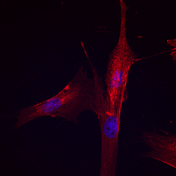
- Integrin beta 1/CD29 in Canine Mesenchymal Stem Cells. Integrin beta 1/CD29 was detected in immersion fixed canine mesenchymal stem cells using Goat Anti-Human/Canine Integrin beta 1/CD29 Antigen Affinity-purified Polyclonal Antibody (Catalog # AF1778) at 10 µg/mL for 3 hours at room temperature. Cells were stained using the NorthernLights™ 557-conjugated Anti-Goat IgG Secondary Antibody (red; Catalog # NL001) and counterstained with DAPI (blue). Specific staining was localized to cell surfaces. View our protocol for Fluorescent ICC Staining of Stem Cells on Coverslips.

- Submitted by
- R&D Systems (provider)
- Main image

- Experimental details
- Integrin beta 1/CD29 in Human Mesenchymal Stem Cells. Integrin beta 1/CD29 was detected in immersion fixed human mesenchymal stem cells using Goat Anti-Human/Canine Integrin beta 1/CD29 Antigen Affinity-purified Polyclonal Antibody (Catalog # AF1778) at 10 µg/mL for 3 hours at room temperature. Cells were stained using the NorthernLights™ 557-conjugated Anti-Goat IgG Secondary Antibody (red; Catalog # NL001) and counterstained with DAPI (blue). Specific staining was localized to cell surfaces. View our protocol for Fluorescent ICC Staining of Stem Cells on Coverslips.

Supportive validation
- Submitted by
- R&D Systems (provider)
- Main image
- Experimental details
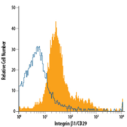
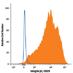
- Detection of Integrin beta 1/CD29 in Canine PBMCs by Flow Cytometry. Canine peripheral blood mononuclear cells (PBMCs) were stained with Goat Anti-Human/Canine Integrin beta 1/CD29 Antigen Affinity-purified Polyclonal Antibody (Catalog # AF1778, filled histogram) or isotype control antibody (Catalog # AB-108-C, open histogram), followed by Phycoerythrin-conjugated Anti-Goat IgG Secondary Antibody (Catalog # F0107).

- Submitted by
- R&D Systems (provider)
- Main image
- Experimental details
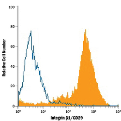
- Detection of Integrin beta 1/CD29 in Human PBMCs by Flow Cytometry. Human peripheral blood mononuclear cells (PBMCs) were stained with Goat Anti-Human/Canine Integrin beta 1/CD29 Antigen Affinity-purified Polyclonal Antibody (Catalog # AF1778, filled histogram) or isotype control antibody (Catalog # AB-108-C, open histogram), followed by Phycoerythrin-conjugated Anti-Goat IgG Secondary Antibody (Catalog # F0107).

- Submitted by
- R&D Systems (provider)
- Main image
- Experimental details
- Detection of Integrin beta 1/CD29 in Canine Mesenchymal Stem Cells by Flow Cytometry. Canine mesenchymal stem cells were stained with Goat Anti-Human/Canine Integrin beta 1/CD29 Antigen Affinity-purified Polyclonal Antibody (Catalog # AF1778, filled histogram) or isotype control antibody (Catalog # AB-108-C, open histogram), followed by Phycoerythrin-conjugated Anti-Goat IgG Secondary Antibody (Catalog # F0107).
