Explore
 Validate
 Learn
Learn Western blot
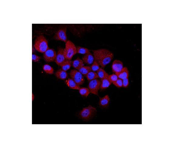
 Immunocytochemistry
Antibody data
- Antibody Data
- Antigen structure
- References [1]
- Comments [0]
- Validations
- Western blot [1]
Submit
Validation data
Reference
Comment
Report error
- Product number
- A02296-3 - Provider product page

- Provider
- Boster Biological Technology
- Product name
- Anti-EEA1 Antibody Picoband™
- Antibody type
- Polyclonal
- Description
- Polyclonal antibody for EEA1 detection. Host: Rabbit.Size: 100μg/vial. Tested applications: WB, ICC/IF, FCM. Reactive species: Human;Mouse;Rat. EEA1 information: Subcellular Localization: Cytoplasm. Early endosome membrane; Peripheral membrane protein.
- Reactivity
- Human, Mouse, Rat
- Host
- Rabbit
- Vial size
- 100μg/vial
- Concentration
- 0.5-1mg/ml, actual concentration vary by lot. Use suggested dilution ratio to decide dilution procedure.
- Storage
- At -20°C for one year. After reconstitution, at 4°C for one month. It can also be aliquoted and stored frozen at -20°C for a longer time. Avoid repeated freezing and thawing.
- Handling
- Add 0.2ml of distilled water will yield a concentration of 500ug/ml.
Submitted references Phagocytosis by endothelial cells inhibits procoagulant activity of platelets of essential thrombocythemia in vitro.
Ji S, Dong W, Qi Y, Gao H, Zhao D, Xu M, Li T, Yu H, Sun Y, Ma R, Shi J, Gao C
Journal of thrombosis and haemostasis : JTH 2020 Jan;18(1):222-233
Journal of thrombosis and haemostasis : JTH 2020 Jan;18(1):222-233
No comments: Submit comment
Supportive validation
- Submitted by
- Boster Biological Technology (provider)
- Main image
- Experimental details
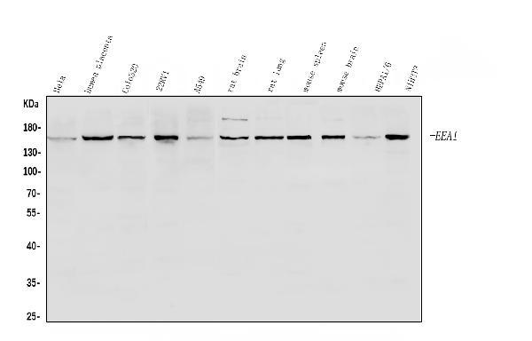
- Western blot analysis of EEA1 using anti-EEA1 antibody (A02296-3). Electrophoresis was performed on a 5-20% SDS-PAGE gel at 70V (Stacking gel) / 90V (Resolving gel) for 2-3 hours. The sample well of each lane was loaded with 30ug of sample under reducing conditions. Lane 1: human Hela whole cell lysates, Lane 2: human placenta tissue lysates, Lane 3: human Colo320 whole cell lysates, Lane 4: human 22RV1 whole cell lysates, Lane 5: human A549 whole cell lysates, Lane 6: rat brain tissue lysates, Lane 7: rat lung tissue lysates, Lane 8: mouse spleen tissue lysates, Lane 9: mouse brain tissue lysates, Lane 10: mouse HEPA1-6 whole cell lysates, Lane 11: mouse NIH/3T3 whole cell lysates. After Electrophoresis, proteins were transferred to a Nitrocellulose membrane at 150mA for 50-90 minutes. Blocked the membrane with 5% Non-fat Milk/ TBS for 1.5 hour at RT. The membrane was incubated with rabbit anti-EEA1 antigen affinity purified polyclonal antibody (Catalog # A02296-3) at 0.5 μg/mL overnight at 4°C, then washed with TBS-0.1%Tween 3 times with 5 minutes each and probed with a goat anti-rabbit IgG-HRP secondary antibody at a dilution of 1:5000 for 1.5 hour at RT. The signal is developed using an Enhanced Chemiluminescent detection (ECL) kit (Catalog # EK1002) with Tanon 5200 system. A specific band was detected for EEA1 at approximately 170KD. The expected band size for EEA1 is at 162KD.
- Additional image