 Explore
 Validate
 Learn
Learn Western blot
Antibody data
- Antibody Data
- Antigen structure
- References [0]
- Comments [0]
- Validations
- Western blot [1]
- Immunohistochemistry [2]
Submit
Validation data
Reference
Comment
Report error
- Product number
- TA319253 - Provider product page

- Provider
- OriGene
- Product name
- Rabbit polyclonal Aurora B pT232 antibody
- Antibody type
- Polyclonal
- Description
- Rabbit polyclonal Aurora B pT232 antibody
- Host
- Rabbit
- Conjugate
- Unconjugated
- Epitope
- AURKB
- Isotype
- IgG
- Antibody clone number
- NULL
- Vial size
- 100 µg
- Concentration
- 1.0 mg/mL
No comments: Submit comment
Supportive validation
- Submitted by
- OriGene (provider)
- Main image
- Experimental details
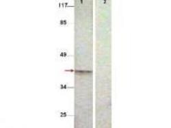
- Western Blot shows detection of Aurora B protein at 39 kDa (predicted band size). All lanes : Aurora B (phospho T232) antibody diluted 1:500. Lane 1 : Extract from COS7 cells treated with Nocodazole (1ug/ml, 16 hrs). Lane 2 : Extract from COS7 cells treated with Nocodazole (1ug/ml, 16 hrs) and with the phosphopeptide immunogen.
- Validation comment
- WB

Supportive validation
- Submitted by
- OriGene (provider)
- Main image
- Experimental details
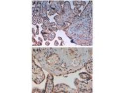
- Immunohistochemistry of Rabbit Anti-AuroraB pT232 Antibody. Tissue: human placenta pH9 (A) at 20x and 40x. Fixation: formalin fixed paraffin embedded. Antigen retrieval: not required. Primary antibody: AuroraB pT232 antibody at 10 ug/mL for 1 h at RT. Secondary antibody: Peroxidase rabbit secondary antibody at 1:10,000 for 45 min at RT. Localization: AuroraB pT232 is cytoplasmic. Staining: AuroraB pT232 as precipitated brown signal with hematoxylin purple nuclear counterstain.
- Validation comment
- IHC

- Submitted by
- OriGene (provider)
- Main image
- Experimental details
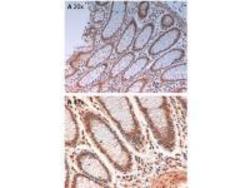
- Immunohistochemistry of Rabbit Anti-AuroraB pT232 Antibody. Tissue: human intestine pH9 (A) at 20x and 40x. Fixation: formalin fixed paraffin embedded. Antigen retrieval: not required. Primary antibody: AuroraB pT232 antibody at 10 ug/mL for 1 h at RT. Secondary antibody: Peroxidase rabbit secondary antibody at 1:10,000 for 45 min at RT. Localization: AuroraB pT232 is cytoplasmic. Staining: AuroraB pT232 as precipitated brown signal with hematoxylin purple nuclear counterstain.
- Validation comment
- IHC
