Explore
 Validate
 Learn
LearnMA5-15321
antibody from Invitrogen Antibodies
Targeting: AURKB
Aik2, AIM-1, ARK2, AurB, IPL1, PPP1R48, STK12, STK5
 Western blot
 ELISA
ELISAAntibody data
- Antibody Data
- Antigen structure
- References [1]
- Comments [0]
- Validations
- Western blot [2]
- Immunocytochemistry [2]
- Other assay [2]
Submit
Validation data
Reference
Comment
Report error
- Product number
- MA5-15321 - Provider product page

- Provider
- Invitrogen Antibodies
- Product name
- Aurora B Monoclonal Antibody (13E8D3)
- Antibody type
- Monoclonal
- Antigen
- Purifed from natural sources
- Description
- MA5-15321 targets AURKB in indirect ELISA, WB applications and shows reactivity with Human samples. The MA5-15321 immunogen is purified recombinant fragment of AURKB expressed in E. Coli.
- Reactivity
- Human
- Host
- Mouse
- Isotype
- IgG
- Antibody clone number
- 13E8D3
- Vial size
- 100 μL
- Concentration
- Conc. Not Determined
- Storage
- Store at 4°C short term. For long term storage, store at -20°C, avoiding freeze/thaw cycles.
Submitted references Klotho Exerts an Emerging Role in Cytokinesis.
Sun CY, Chou CY, Hsieh YY, Lo KC, Liou YL, Chen YH
Genes 2020 Sep 4;11(9)
Genes 2020 Sep 4;11(9)
No comments: Submit comment
Supportive validation
- Submitted by
- Invitrogen Antibodies (provider)
- Main image
- Experimental details
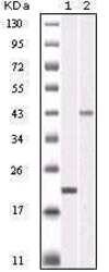
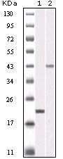
- Western blot analysis of AURKB using a AURKB monoclonal antibody (Product # MA5-15321) against a truncated AURKB recombinant protein (1) and SKN-SH cell lysate (2).
- Submitted by
- Invitrogen Antibodies (provider)
- Main image
- Experimental details
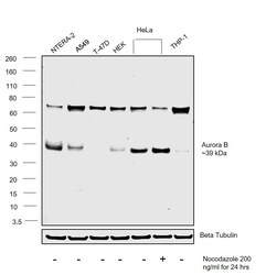
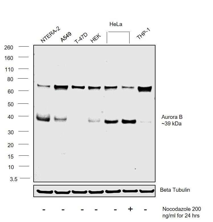
- Western blot was performed using Aurora B Monoclonal Antibody (13E8D3) (Product # MA5-15321) and a 39 kDa band corresponding to Aurora B was observed across cell lines tested and increased upon Nocodazole treatment in HeLa. Whole cell extracts (30 µg lysate) of NTERA-2 (Lane 1), A-549 (Lane 2), T-47D (Lane 3), HEK (Lane 4), HeLa (Lane 5), HeLa treated with Nocodazole (200ng/ml for 24 hours (Lane 6) and THP-1 (Lane 7) were electrophoresed using Novex® NuPAGE® 12 % Bis-Tris gel (Product # NP0342BOX). Resolved proteins were then transferred onto a nitrocellulose membrane (Product # IB23001) by iBlot® 2 Dry Blotting System (Product # IB21001). The blot was probed with the primary antibody (1:1000 dilution) and detected by chemiluminescence with Goat anti-Mouse IgG (H+L) Superclonal™ Recombinant Secondary Antibody, HRP (Product # A28177, 1:4000 dilution) using the iBright FL 1000 (Product # A32752). Chemiluminescent detection was performed using Novex® ECL Chemiluminescent Substrate Reagent Kit (Product # WP20005)..
Supportive validation
- Submitted by
- Invitrogen Antibodies (provider)
- Main image
- Experimental details
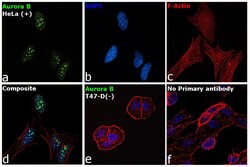
- Immunofluorescence analysis of Aurora B was performed using 70% confluent log phase HeLa and T-47D cells. The cells were fixed with 4% Paraformaldehyde for 10 minutes, permeabilized with 0.1% Triton™ X-100 for 10 minutes, and blocked with 2% BSA for 10 minutes at room temperature. The cells were labeled with Aurora B Monoclonal Antibody (13E8D3) (Product # MA5-15321) at 1:200 dilution in 0.1% BSA, incubated at 4 degree celsius overnight and then labeled with Goat anti-Mouse IgG (H+L) Superclonal™ Secondary Antibody, Alexa Fluor® 488 conjugate (Product # A28175, 1:2000 dilution) for 45 minutes at room temperature (Panel a: Green). Nuclei (Panel b: Blue) were stained with SlowFade® Gold Antifade Mountant with DAPI (Product # S36938). F-actin (Panel c: Red) was stained with Rhodamine Phalloidin (Product # R415, 1:300). Panel d represents the merged image showing plasma membrane localization. Panel e represents T-47D cells having low expression of Aurora B. Panel f represents control cells with no primary antibody to assess background. The images were captured at 60X magnification.

- Submitted by
- Invitrogen Antibodies (provider)
- Main image

- Experimental details
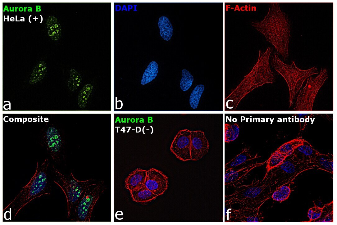
- Immunofluorescence analysis of Aurora B was performed using 70% confluent log phase HeLa and T-47D cells. The cells were fixed with 4% Paraformaldehyde for 10 minutes, permeabilized with 0.1% Triton™ X-100 for 10 minutes, and blocked with 2% BSA for 10 minutes at room temperature. The cells were labeled with Aurora B Monoclonal Antibody (13E8D3) (Product # MA5-15321) at 1:200 dilution in 0.1% BSA, incubated at 4 degree celsius overnight and then labeled with Goat anti-Mouse IgG (H+L) Superclonal™ Secondary Antibody, Alexa Fluor® 488 conjugate (Product # A28175, 1:2000 dilution) for 45 minutes at room temperature (Panel a: Green). Nuclei (Panel b: Blue) were stained with SlowFade® Gold Antifade Mountant with DAPI (Product # S36938). F-actin (Panel c: Red) was stained with Rhodamine Phalloidin (Product # R415, 1:300). Panel d represents the merged image showing plasma membrane localization. Panel e represents T-47D cells having low expression of Aurora B. Panel f represents control cells with no primary antibody to assess background. The images were captured at 60X magnification.
Supportive validation
- Submitted by
- Invitrogen Antibodies (provider)
- Main image
- Experimental details
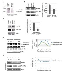
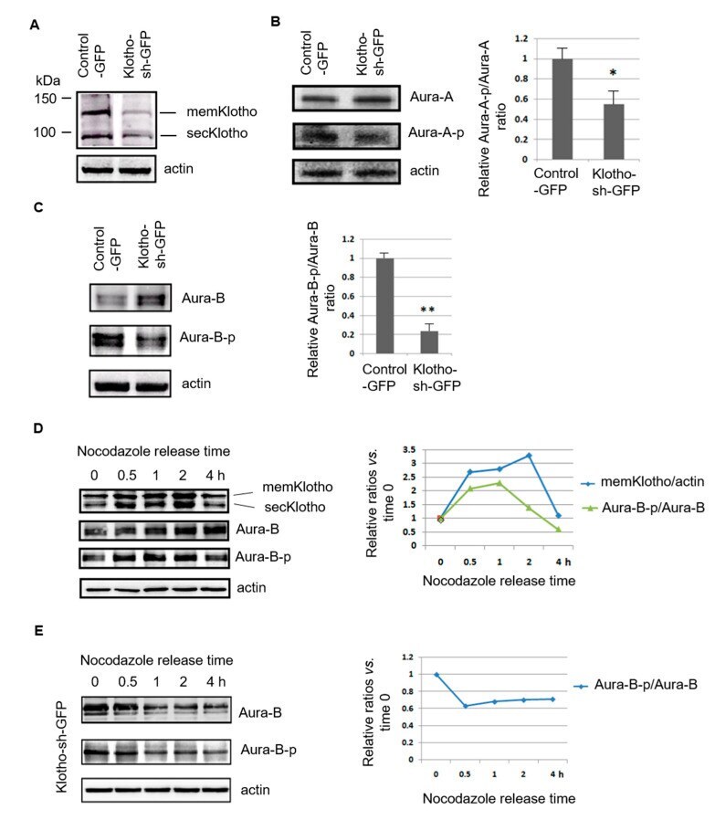
- Figure 6 Klotho depletion impairs Aurora kinase activation. ( A - C ) Immunoblot analysis of Klotho, and Aurora kinase A and B with the protein extracts of transfected HeLa cells, respectively. ( D , E ) Immunoblot analysis of Klotho and Aurora kinase B with the protein extracts of HeLa cells treated with nocodazole for 16 h, respectively. Each treatment for ( B , C ) was repeated in triplicate, and the statistic results are plotted (means +- SEM; one-way ANOVA; * p < 0.05; ** p < 0.01; KL--Klotho; Aura A--Aurora kinase A; Aura B--Aurora kinase B).
- Submitted by
- Invitrogen Antibodies (provider)
- Main image
- Experimental details
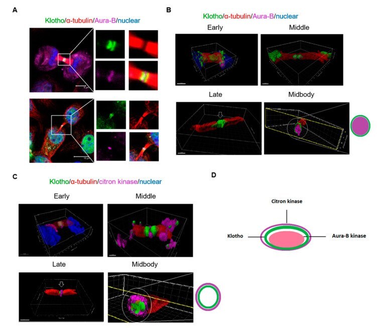
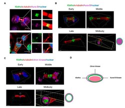
- Figure 7 3D reconstruction of the cytokinesis bridge. Intrinsic Klotho, Aurora kinase B, and citron kinase of cultured HeLa cells analyzed by immunofluorescence. ( A ) Results of immunofluorescence staining for Klotho and Aurora kinase B. ( B ) 3D structure of Klotho and Aurora kinase B in the cytokinesis bridge. ( C ) 3D structure of Klotho and citron kinase in the cytokinesis bridge. ( D ) Midbody architecture related with Klotho, Aurora kinase B, and citron kinase (microscopy: x400).