Explore
 Validate
 Learn
LearnAP09251PU-N
antibody from Acris Antibodies GmbH
Targeting: AURKB
Aik2, AIM-1, ARK2, AurB, IPL1, PPP1R48, STK12, STK5
 Western blot
 ELISA
ELISAAntibody data
- Antibody Data
- Antigen structure
- References [0]
- Comments [0]
- Validations
- Western blot [1]
- Immunohistochemistry [2]
Submit
Validation data
Reference
Comment
Report error
- Product number
- AP09251PU-N - Provider product page

- Provider
- Acris Antibodies GmbH
- Proper citation
- Acris Antibodies GmbH Cat#AP09251PU-N, RRID:AB_2035157
- Product name
- anti Aurora kinase B pThr232
- Antibody type
- Polyclonal
- Antigen
- Synthetic peptide corresponding aa 223-234 of Human Aurora Kinase B protein
- Reactivity
- Human, Mouse, Rat, Bovine, Canine, Porcine
- Host
- Rabbit
- Isotype
- IgG
- Vial size
- 0.1 mg
- Concentration
- 1.0 mg/ml (by UV absorbance at 280 nm)
No comments: Submit comment
Supportive validation
- Submitted by
- Acris Antibodies GmbH (provider)
- Main image
- Experimental details
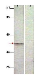
- Western Blot shows detection of Aurora B protein at 39 kDa (predicted band size). All Lanes : Aurora B (phospho T232) antibody diluted 1/500. Lane 1 : Extract from COS7 cells treated with Nocodazole (1 µg/ml, 16 hrs). Lane 2 : Extract from COS7 cells treated with Nocodazole (1 µg/ml, 16 hrs) and with the phosphopeptide immunogen.

Supportive validation
- Submitted by
- Acris Antibodies GmbH (provider)
- Main image
- Experimental details
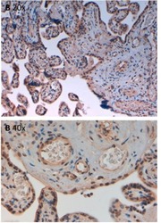
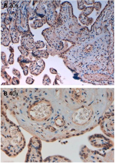
- Immunohistochemistry of Rabbit Anti-AuroraB pT232 Antibody. Tissue: human placenta pH9 (A) at 20x and 40x. Fixation: formalin fixed paraffin embedded. Antigen retrieval: not required. Primary antibody: AuroraB pT232 antibody at 10 µg/mL for 1 h at RT. Secondary antibody: Peroxidase rabbit secondary antibody at 1:10,000 for 45 min at RT. Localization: AuroraB pT232 is cytoplasmic. Staining: AuroraB pT232 as precipitated brown signal with hematoxylin purple nuclear counterstain.
- Submitted by
- Acris Antibodies GmbH (provider)
- Main image

- Experimental details
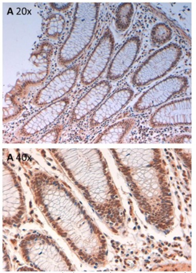
- Immunohistochemistry of Rabbit Anti-AuroraB pT232 Antibody. Tissue: human intestine pH9 (A) at 20x and 40x. Fixation: formalin fixed paraffin embedded. Antigen retrieval: not required. Primary antibody: AuroraB pT232 antibody at 10 µg/mL for 1 h at RT. Secondary antibody: Peroxidase rabbit secondary antibody at 1:10,000 for 45 min at RT. Localization: AuroraB pT232 is cytoplasmic. Staining: AuroraB pT232 as precipitated brown signal with hematoxylin purple nuclear counterstain.