 Explore
 Validate
 Learn
Learn Western blot
Antibody data
- Antibody Data
- Antigen structure
- References [0]
- Comments [0]
- Validations
- Western blot [5]
- Immunocytochemistry [1]
- Immunohistochemistry [3]
- Other assay [1]
Submit
Validation data
Reference
Comment
Report error
- Product number
- PA1-31019 - Provider product page

- Provider
- Invitrogen Antibodies
- Product name
- DDX5 Polyclonal Antibody
- Antibody type
- Polyclonal
- Antigen
- Synthetic peptide
- Description
- Recommended positive controls: The peptide used to generate this antibody is available for purchase (GTX10261-PEP).. Store product as a concentrated solution. Centrifuge briefly prior to opening the vial.
- Reactivity
- Human, Mouse
- Host
- Goat
- Isotype
- IgG
- Vial size
- 100 µg
- Concentration
- 0.5 mg/mL
- Storage
- Store at 4°C short term. For long term storage, store at -20°C, avoiding freeze/thaw cycles.
No comments: Submit comment
Supportive validation
- Submitted by
- Invitrogen Antibodies (provider)
- Main image

- Experimental details
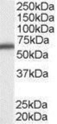
- Western blot analysis of DDX5 in A431 cell lysate (RIPA buffer, 35 µg total protein per lane) using a DDX5 polyclonal antibody (Product # PA1-31019) at a dilution of 0.1 µg/mL following incubation for 1 hour and detected using chemiluminescence.

- Submitted by
- Invitrogen Antibodies (provider)
- Main image
- Experimental details
- Western blot analysis of DDX5 in A431 cell lysate (RIPA buffer, 35 µg total protein per lane) using a DDX5 polyclonal antibody (Product # PA1-31019) at a dilution of 0.5 µg/mL following incubation for 1 hour and detected using chemiluminescence.

- Submitted by
- Invitrogen Antibodies (provider)
- Main image
- Experimental details
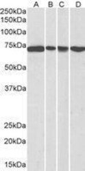
- Western Blot analysis of DDX5 was performed by loading 35 µg (in RIPA buffer) of NIH-3T3 (A), HeLa (B), A431 (C) and Jurkat (D) nuclear lysates. Proteins were transferred to a membrane and probed with a DDX5 Polyclonal Antibody (Product # PA1-31019) at a dilution of 0.3 µg/mL.

- Submitted by
- Invitrogen Antibodies (provider)
- Main image
- Experimental details
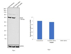
- Knockdown of DDX5 was achieved by transfecting HeLa with DDX5 specific siRNAs (Silencer® select Product # s4008, Product # s4009). Western blot analysis (Fig. a) was performed using whole cell extracts from the DDX5 knockdown cells (lane 3), non-specific scrambled siRNA transfected cells (lane 2) and untransfected cells (lane 1). The blot was probed with DDX5 Polyclonal Antibody (Product # PA1-31019, 1:1000 dilution) and Rabbit anti-goat IgG (H+L) Recombinant Secondary Antibody, HRP (Product # A27014, 0.25µg/ml, 1:4000 dilution). Densitometric analysis of this western blot is shown in histogram (Fig. b). Decrease in signal upon siRNA mediated knock down confirms that antibody is specific to DDX5.

- Submitted by
- Invitrogen Antibodies (provider)
- Main image
- Experimental details
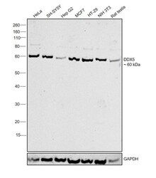
- Western blot was performed using Anti-DDX5 Polyclonal Antibody (Product # PA1-31019) and a 60 kDa band corresponding to DDX5 was observed across cell lines and tissues tested. Modified whole cell extracts (1% SDS) (30 µg lysate) of HeLa (Lane 1), SH-SY5Y (Lane 2),Hep G2 (Lane 3), MCF7 (Lane 4), HT-29 (Lane 5), NIH/3T3 (Lane 6) and Rat testis (Lane 7) were electrophoresed using Novex® NuPAGE® 4-12 % Bis-Tris gel (Product # NP0322BOX). Resolved proteins were then transferred onto a nitrocellulose membrane (Product # IB23001) by iBlot® 2 Dry Blotting System (Product # IB21001). The blot was probed with the primary antibody (1:1000 dilution) and detected by chemiluminescence with Rabbit Anti-Goat IgG (H+L), Superclonal™ Recombinant Secondary Antibody, HRP (Product # A27014, 1:4000 dilution) using the iBright FL 1000 (Product # A32752). Chemiluminescent detection was performed using Novex® ECL Chemiluminescent Substrate Reagent Kit (Product # WP20005).

Supportive validation
- Submitted by
- Invitrogen Antibodies (provider)
- Main image

- Experimental details
- Immunofluorescence analysis of DDX5 was performed using 70% confluent log phase MCF7 cells. The cells were fixed with 4% paraformaldehyde for 10 minutes, permeabilized with 0.1% Triton™ X-100 for 15 minutes, and blocked with 2% BSA for 1 hour at room temperature. The cells were labeled with DDX5 Polyclonal Antibody (Product # PA1-31019) at 1:100 dilution in 0.1% BSA, incubated at 4 degree Celsius overnight and then labeled with Rabbit anti-Goat IgG (H+L) Recombinant Secondary Antibody, Alexa Fluor® 488 conjugate (Product # A-11078) at a dilution of 1:2000 for 45 minutes at room temperature (Panel a: green). Nuclei (Panel b: blue) were stained with SlowFade® Gold Antifade Mountant with DAPI (Product # S36938). F-actin (Panel c: red) was stained with Rhodamine Phalloidin (Product # R415, 1:300). Panel d represents the merged image showing localization to the nucleus. Panel e represents control cells with no primary antibody to assess background. The images were captured at 60X magnification.

Supportive validation
- Submitted by
- Invitrogen Antibodies (provider)
- Main image
- Experimental details
- Immunohistochemistry (Paraffin) analysis of DDX5 in human breast tissue using DDX5 Polyclonal Antibody (Product # PA1-31019) at a dilution of 2.5 µg/mL. Antigen retrieval : citrate buffer pH 6.

- Submitted by
- Invitrogen Antibodies (provider)
- Main image

- Experimental details
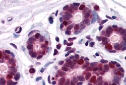
- Immunohistochemistry (Paraffin) analysis of DDX5 in human prostate tissue using DDX5 Polyclonal Antibody (Product # PA1-31019) at a dilution of 2.5 µg/mL. Antigen retrieval : citrate buffer pH 6.

- Submitted by
- Invitrogen Antibodies (provider)
- Main image
- Experimental details
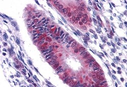
- Immunohistochemistry (Paraffin) analysis of DDX5 in human uterus tissue using DDX5 Polyclonal Antibody (Product # PA1-31019) at a dilution of 2.5 µg/mL. Antigen retrieval : citrate buffer pH 6.

Supportive validation
- Submitted by
- Invitrogen Antibodies (provider)
- Main image

- Experimental details
- RNA Immunoprecipitation (RIP) assay of endogenous DDX5 protein using Anti-DDX5 Antibody: RIP assay was performed using Anti-DDX5 Polyclonal Antibody (Product # PA1-31019, 5 ug) on whole cell lysate from HeLa cells. Normal IgG was used as a negative IP control. RNA purified by RiboPure™ RNA Purification Kit (Product # AM1924) was analyzed by RT-PCR using the Power SYBR® Green RNA-to-CT™ 1-Step Kit (Product # 4389986) with the primers pairs over U1 snRNA, ACTB, MALAT non-coding RNA and 5S rRNA. Data is presented as fold enrichment of the antibody signal versus the negative control IgG using the comparative CT method.
