 Explore
 Validate
 Learn
Learn Western blot
Antibody data
- Antibody Data
- Antigen structure
- References [0]
- Comments [0]
- Validations
- Western blot [4]
- Immunohistochemistry [3]
Submit
Validation data
Reference
Comment
Report error
- Product number
- PA5-18260 - Provider product page

- Provider
- Invitrogen Antibodies
- Product name
- DDX5 Polyclonal Antibody
- Antibody type
- Polyclonal
- Antigen
- Synthetic peptide
- Description
- This antibody is predicted to react with canine, mouse and rat based on sequence homology. This antibody is tested in Peptide ELISA: antibody detection limit dilution 2,000.
- Reactivity
- Human, Mouse
- Host
- Goat
- Isotype
- IgG
- Vial size
- 100 µg
- Concentration
- 0.5 mg/mL
- Storage
- -20° C, Avoid Freeze/Thaw Cycles
No comments: Submit comment
Supportive validation
- Submitted by
- Invitrogen Antibodies (provider)
- Main image

- Experimental details
- Western blot analysis of DDX5 using a DDX5 polyclonal antibody (Product # PA5-18260) at a concentration of 0.5 µg/mL. A431(A), HeLa(B), HepG2(C), Jurkat(D) and NIH3T3 (E) lysate (35 µg protein in RIPA buffer). Primary incubation was 1 hour. Detected by chemiluminescence.

- Submitted by
- Invitrogen Antibodies (provider)
- Main image

- Experimental details
- Western Blot staining of A431 cell lysate using Product # PA5-18260 at a concentration of 0.5 µg/mL, the primary antibody incubation was 1 hour and the detection method was chemiluminescence.

- Submitted by
- Invitrogen Antibodies (provider)
- Main image

- Experimental details
- Western blot analysis was performed on modified whole cell extracts (1% SDS) (30 µg lysate) of A-431 (Lane 1), IMR-32 (Lane 2), HeLa (Lane 3), K-562 (Lane 4), HT-29 (Lane 5), MOLT-4 (Lane 6), Jurkat (Lane 7), Hep G2 (Lane 8), MCF7 (Lane 9), SH-SY5Y (Lane 10) and NIH/3T3 (Lane 11). The blot was probed with Anti-DDX5 Polyclonal Antibody (Product # PA5-18260, 1 µg/ml) and detected by Rabbit anti-Goat IgG (H+L) Superclonal™ Secondary Antibody, HRP (Product # A27014, 0.25 µg/ml, 1:4000 dilution). A 69 kDa band corresponding to DDX5 was detected across the cell lines tested.

- Submitted by
- Invitrogen Antibodies (provider)
- Main image

- Experimental details
- Knockdown of DDX5 was achieved by transfecting HeLa cells with DDX5 specific siRNAs (Silencer® select Product # s4009, s4008). Western blot analysis (Fig. a) was performed using modified whole cell extracts (0.1% SDS) from the DDX5 knockdown cells (lane 3), non-specific scrambled siRNA transfected cells (lane 2) and untransfected cells (lane 1). The blots were probed with DDX5 Polyclonal Antibody (Product # PA5-48035, 1 µg/ml) and Rabbit anti-Goat IgG (H+L) Superclonal™ Secondary Antibody, HRP (Product # A27014, 0.25 µg/ml, 1:4000 dilution). Densitometric analysis of this western blot is shown in histogram (Fig. b). Decrease in signal upon siRNA mediated knock down confirms that antibody is specific to DDX5.

Supportive validation
- Submitted by
- Invitrogen Antibodies (provider)
- Main image

- Experimental details
- Immunohistochemical analysis of DDX5 in Human Breast using a DDX5 monoclonal antibody (Product #PA5-18260) at 2.5 µg/mL. The Human Breast tissue section was paraffin embeded and detected using steamed antigen retrieval with citrate buffer pH 6, AP-staining.

- Submitted by
- Invitrogen Antibodies (provider)
- Main image

- Experimental details
- Immunohistochemical analysis of DDX5 in Human Uterus using a DDX5 monoclonal antibody (Product #PA5-18260) at 2.5 µg/mL. The Human Uterus tissue section was paraffin embeded and detected using steamed antigen retrieval with citrate buffer pH 6, AP-staining.
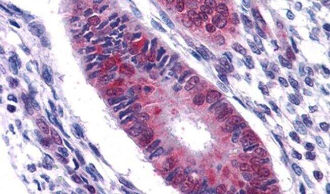
- Submitted by
- Invitrogen Antibodies (provider)
- Main image

- Experimental details
- Immunohistochemichal analysis of DDX5 in Human Prostate using a DDX5 polyclonal antibody (Product # PA5-18260) at a concentration of 2.5 µg/mL. The sample was paraffin embedded, and heat treated antigen retrieval was used to detect the target with AP staining.
