Explore
 Validate
 Learn
Learn Western blot
Antibody data
- Antibody Data
- Antigen structure
- References [0]
- Comments [0]
- Validations
- Western blot [1]
- Immunohistochemistry [1]
Submit
Validation data
Reference
Comment
Report error
- Product number
- TA319363 - Provider product page

- Provider
- OriGene
- Product name
- Rabbit polyclonal anti-iASSP antibody
- Antibody type
- Polyclonal
- Description
- Rabbit polyclonal anti-iASSP antibody
- Host
- Rabbit
- Conjugate
- Unconjugated
- Epitope
- PPP1R13L
- Isotype
- IgG
- Antibody clone number
- NULL
- Vial size
- 100 µg
- Concentration
- 1.0 mg/mL
No comments: Submit comment
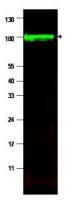
Supportive validation
- Submitted by
- OriGene (provider)
- Main image
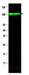
- Experimental details
- WB using Anti-iASPP antibody shows detection of a band at ~100 kDa (arrowhead) corresponding to isoform 1 of iASPP in MCF7 whole cell lysates. Preincubation with immunizing peptide blocks specific band staining (data not shown). Approximately 35 ug of lysate was separated by SDS-PAGE. After blocking, the membrane was probed with the primary antibody diluted to 1:1,500. The membrane was washed and reacted with a 1:10,000 dilution of IRDye800 conjugated Gt-a-Rabbit IgG [H&L].
- Validation comment
- WB
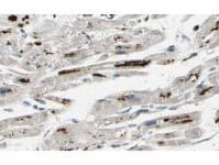
Supportive validation
- Submitted by
- OriGene (provider)
- Main image
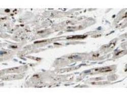
- Experimental details
- Affinity Purified anti-iASPP antibody shows strong cytoplasmic and membranous staining of myocytes in human heart tissue. Tissue was formalin-fixed and paraffin embedded. Brown color indicates presence of protein, blue color shows cell nuclei. Personal Communication, Kenneth Wester, www.proteinatlas.org, Uppsala, Sweden.
- Validation comment
- IHC