Explore
 Validate
 Learn
Learn Western blot
 ELISA
ELISA Immunohistochemistry
Antibody data
- Antibody Data
- Antigen structure
- References [0]
- Comments [0]
- Validations
- Western blot [1]
- Immunohistochemistry [1]
Submit
Validation data
Reference
Comment
Report error
- Product number
- LS-C745300 - Provider product page

- Provider
- LSBio
- Product name
- PPP1R13L / iASPP Antibody LS-C745300
- Antibody type
- Polyclonal
- Description
- Affinity purified
- Reactivity
- Human
- Host
- Rabbit
- Isotype
- IgG
- Storage
- Store vial at -20°C or below prior to opening. Dilute 1:10 to minimize loss. Store the vial at -20°C or below after dilution. Avoid freeze-thaw cycles.
No comments: Submit comment
Enhanced validation
- Submitted by
- LSBio (provider)
- Enhanced method
- Genetic validation
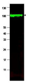
- Main image

- Experimental details
- Western blot using the affinity purified anti-iASPP antibody shows detection of a band at ~100 kDa (arrowhead) corresponding to isoform 1 of iASPP in MCF7 whole cell lysates. Preincubation with immunizing peptide blocks specific band staining (data not shown). Approximately 35 ug of lysate was separated by 4-20% Tris Glycine SDS-PAGE. After blocking, the membrane was probed with the primary antibody diluted to 1:1,500 in 5% BLOTTO/PBS overnight at 4°C. The membrane was washed and reacted with a 1:10,000 dilution of conjugated Gt-a-Rabbit IgG [H&L] for 45 min at room temperature (800 nm channel, green). Molecular weight estimation was made by comparison to prestained MW markers. Fluorescence image was captured using the Odyssey Infrared Imaging System developed by LI-COR. Other detection systems will yield similar results.
Supportive validation
- Submitted by
- LSBio (provider)
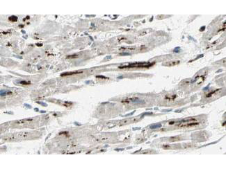
- Main image
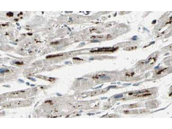
- Experimental details
- Affinity Purified anti-iASPP antibody shows strong cytoplasmic and membranous staining of myocytes in human heart tissue. Tissue was formalin-fixed and paraffin embedded. Brown color indicates presence of protein, blue color shows cell nuclei.