Explore
 Validate
 Learn
Learn Western blot
 ELISA
ELISAAntibody data
- Antibody Data
- Antigen structure
- References [0]
- Comments [0]
- Validations
- Western blot [1]
- Immunohistochemistry [1]
Submit
Validation data
Reference
Comment
Report error
- Product number
- 600-401-926 - Provider product page

- Provider
- Invitrogen Antibodies
- Product name
- iASSP Polyclonal Antibody
- Antibody type
- Polyclonal
- Antigen
- Synthetic peptide
- Reactivity
- Human
- Host
- Rabbit
- Isotype
- IgG
- Vial size
- 100 µg
- Concentration
- 1.1 mg/mL
- Storage
- -20° C, Avoid Freeze/Thaw Cycles
No comments: Submit comment
Supportive validation
- Submitted by
- Invitrogen Antibodies (provider)
- Main image
- Experimental details
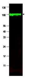
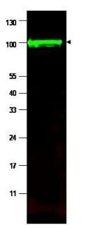
- Western blot using Rocklands affinity purified anti-iASPP antibody shows detection of a band at ~100 kDa (arrowhead) corresponding to isoform 1 of iASPP in MCF7 whole cell lysates. Preincubation with immunizing peptide blocks specific band staining (data not shown). Approximately 35 µg of lysate was separated by 4-20% Tris Glycine SDS-PAGE. After blocking, the membrane was probed with the primary antibody diluted to 1:1,500 in 5% BLOTTO/PBS overnight at 4°C. The membrane was washed and reacted with a 1:10,000 dilution of IRDye800 conjugated Gt-a-Rabbit IgG [H&L] (611-132-122) for 45 min at room temperature (800 nm channel, green). Molecular weight estimation was made by comparison to prestained MW markers. IRDye800 fluorescence image was captured using the Odyssey® Infrared Imaging System developed by LI-COR. IRDye is a trademark of LI-COR, Inc. Other detection systems will yield similar results.
Supportive validation
- Submitted by
- Invitrogen Antibodies (provider)
- Main image
- Experimental details
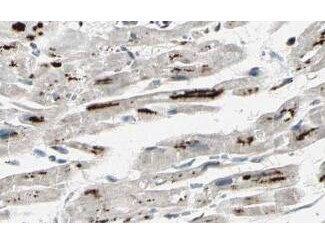
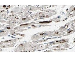
- Rocklands Affinity Purified anti-iASPP antibody shows strong cytoplasmic and membranous staining of myocytes in human heart tissue. Tissue was formalin-fixed and paraffin embedded. Brown color indicates presence of protein, blue color shows cell nuclei. Personal Communication, Kenneth Wester, www.proteinatlas.org, Uppsala, Sweden.