 Explore
 Validate
 Learn
Learn Western blot
Antibody data
- Antibody Data
- Antigen structure
- References [0]
- Comments [0]
- Validations
- Western blot [1]
- Immunohistochemistry [9]
- Flow cytometry [1]
Submit
Validation data
Reference
Comment
Report error
- Product number
- NBP2-27401 - Provider product page

- Provider
- Novus Biologicals
- Product name
- Mouse Monoclonal HMGB1/HMG-1 Antibody
- Antibody type
- Monoclonal
- Description
- Protein G purified. This antibody recognizes both the box A domain (amino acids 9-79) and full length HMGB1 protein.
- Reactivity
- Human, Mouse
- Host
- Mouse
- Isotype
- IgG
- Vial size
- 0.1 mg
- Concentration
- 0.5 mg/ml
- Storage
- Store at 4C short term. Aliquot and store at -20C long term. Avoid freeze-thaw cycles.
No comments: Submit comment
Supportive validation
- Submitted by
- Novus Biologicals (provider)
- Main image
- Experimental details
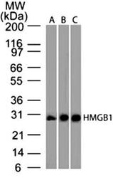
- Western Blot: HMGB1/HMG-1 Antibody (19N15F4) [NBP2-27401] - Analysis of HMGB1 protein on ( A) Full-length human HMGB1 protein (B) human Jurkat cell lysate and (C) mouse NIH 3T3 cell lysate using HMGB1 antibody (clone 19N15F4) at 2 ug/ml concentration. Goat anti-mouse IgG HRP secondary antibody and PicoTect ECL substrate solution were used in this assay.

Supportive validation
- Submitted by
- Novus Biologicals (provider)
- Main image
- Experimental details
- Immunohistochemistry-Paraffin: HMGB1/HMG-1 Antibody (19N15F4) [NBP2-27401] - Analysis of HMGB1 protein in a section of human breast normal tissue using HMGB1 antibody (clone 19N15F4) at a concentration of 5 ug/ml. The ductal /acinar epithelial cells in the breast tissue section depicted strong nuclear expression with some cytoplasmic positivity. The myoepithelial cells and few cells of the intra-lobular connective tissue showed relatively weak nuclear positivity for HMGB1.

- Submitted by
- Novus Biologicals (provider)
- Main image

- Experimental details
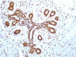
- Immunohistochemistry-Paraffin: HMGB1/HMG-1 Antibody (19N15F4) [NBP2-27401] - Analysis of HMGB1 protein in a section of human small intestinal cancer tissue using HMGB1 antibody (clone 19N15F4) at a concentration of 5 ug/ml. Intense nuclear immunopositivity of HMGB1 was observed in cancer cells and the cells of tumor stroma as well as the adjacent normal crypts. Some glandular cells in the crypts developed bother nuclear and cytoplasmic staining.

- Submitted by
- Novus Biologicals (provider)
- Main image
- Experimental details
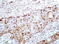
- Immunohistochemistry-Paraffin: HMGB1/HMG-1 Antibody (19N15F4) [NBP2-27401] - Analysis of HMGB1 protein in a tissue section of human esophageal squamous cell carcinoma (SCC) using HMGB1 antibody (clone 19N15F4) at a concentration of 5 ug/ml. Strong nuclear along with weak cytoplasmic immunopositivity for HMGB1 was observed in SCC cells and the tumor stroma cells developed relatively weak staining for this target.

- Submitted by
- Novus Biologicals (provider)
- Main image

- Experimental details
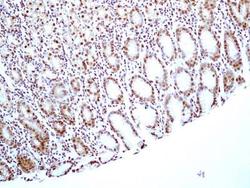
- Immunohistochemistry-Paraffin: HMGB1/HMG-1 Antibody (19N15F4) [NBP2-27401] - Analysis of HMGB1 protein in a tissue section of normal human colon using HMGB1 antibody (clone 19N15F4) at a concentration of 5 ug/ml. Almost all the cells of colon's mucosal layer showed nuclear positivity for HMGB1 but the cells of the columnar epithelial cells on the absorptive surface developed cytoplasmic staining also.

- Submitted by
- Novus Biologicals (provider)
- Main image

- Experimental details
- Immunohistochemistry-Paraffin: HMGB1/HMG-1 Antibody (19N15F4) [NBP2-27401] - Analysis of HMGB1 protein in a tissue section of human bladder transitional cell carcinoma (TCC) / urothelial cell carcinoma (UCC) using HMGB1 antibody (clone 19N15F4) at a concentration of 5 ug/ml. Almost all the cancer cells and the cells of tumor stroma developed strong/specific nuclear HMGB1 immunopositivity.

- Submitted by
- Novus Biologicals (provider)
- Main image

- Experimental details
- Immunohistochemistry-Paraffin: HMGB1/HMG-1 Antibody (19N15F4) [NBP2-27401] - Analysis of HMGB1 protein in a tissue section of normal human brain using HMGB1 antibody (clone 19N15F4) at a concentration of 5 ug/ml. The representative image shows an overall strong nuclear HMGB1 immunopositivity with weak to negligible cytoplasmic staining in brain cells.

- Submitted by
- Novus Biologicals (provider)
- Main image
- Experimental details
- Immunohistochemistry-Paraffin: HMGB1/HMG-1 Antibody (19N15F4) [NBP2-27401] - Analysis of HMGB1 protein in a section of normal human stomach tissue using HMGB1 antibody (clone 19N15F4) at a concentration of 5 ug/ml. The cells of the glandular stomach showed specific nuclear expression for HMGB1.

- Submitted by
- Novus Biologicals (provider)
- Main image

- Experimental details
- Immunohistochemistry-Paraffin: HMGB1/HMG-1 Antibody (19N15F4) [NBP2-27401] - Formalin-fixed, paraffin-embedded human skin stained with HMGB1 antibody at 5 ug/ml

- Submitted by
- Novus Biologicals (provider)
- Main image

- Experimental details
- Immunohistochemistry-Paraffin: HMGB1/HMG-1 Antibody (19N15F4) [NBP2-27401] - IHC-P detection of HMGB1 protein in formalin-fixed, decalcified, paraffin embedded tissue sections from the paws of mouse (collagen-induced arthritis model) using HMGB1 antibody (clone 19N15F4) at a concentration of 5 ug/ml [Image courtesy of Dr Ulf Andersson, Karolinska]

Supportive validation
- Submitted by
- Novus Biologicals (provider)
- Main image
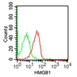
- Experimental details
- Flow Cytometry: HMGB1/HMG-1 Antibody (19N15F4) [NBP2-27401] - Intracellular analysis using HMGB1 antibody. Human Jurkat cells were probed using 0.5 ug of HMGB1 antibody (red) and 0.5 ug of isotype control antibody (green).
