Explore
 Validate
 Learn
Learn Western blot
 Immunoprecipitation
Antibody data
- Antibody Data
- Antigen structure
- References [1]
- Comments [0]
- Validations
- Western blot [3]
- Immunohistochemistry [12]
- Flow cytometry [1]
Submit
Validation data
Reference
Comment
Report error
- Product number
- NBP2-27396 - Provider product page

- Provider
- Novus Biologicals
- Product name
- Mouse Monoclonal HMGB1/HMG-1 Antibody
- Antibody type
- Monoclonal
- Description
- Protein G purified. This antibody recognizes both A box domain (amino acids 9-79) and full length HMGB1 protein.
- Reactivity
- Human, Mouse
- Host
- Mouse
- Isotype
- IgG
- Vial size
- 0.1 mg
- Concentration
- 1.0 mg/ml
- Storage
- Store at 4C short term. Aliquot and store at -20C long term. Avoid freeze-thaw cycles.
Submitted references HMGB1 accelerates alveolar epithelial repair via an IL-1β- and αvβ6 integrin-dependent activation of TGF-β1.
Pittet JF, Koh H, Fang X, Iles K, Christiaans S, Anjun N, Wagener BM, Park DW, Zmijewski JW, Matthay MA, Roux J
PloS one 2013;8(5):e63907
PloS one 2013;8(5):e63907
No comments: Submit comment
Supportive validation
- Submitted by
- Novus Biologicals (provider)
- Main image

- Experimental details
- Simple Western: HMGB1/HMG-1 Antibody (19N10B7) [NBP2-27396] - Simple Western lane view shows a specific band for HMGB1 in 1.0 mg/ml of HeLa lysate. This experiment was performed under reducing conditions using the 12-230kDa separation system.

- Submitted by
- Novus Biologicals (provider)
- Main image

- Experimental details
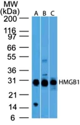
- Western Blot: HMGB1/HMG-1 Antibody (19N10B7) [NBP2-27396] - Analysis using 2 ug/ml concentration of HMGB1 antibody (clone 19N10B7) on (A) Full-length human HMGB1 protein, (B) Human Jurkat cell lysate and (C) Mouse NIH 3T3 cell lysate. Goat anti-mouse Ig HRP secondary antibody and PicoTect ECL substrate solution was used for this test.
- Submitted by
- Novus Biologicals (provider)
- Main image
- Experimental details
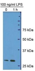
- Western Blot: HMGB1/HMG-1 Antibody (19N10B7) [NBP2-27396] - Hepatocyte protein lysate at 1:1000 4C overnight. Image from verified customer review.

Supportive validation
- Submitted by
- Novus Biologicals (provider)
- Main image

- Experimental details
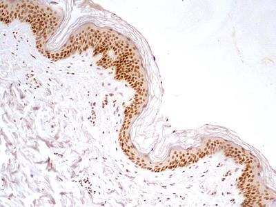
- Immunohistochemistry-Paraffin: HMGB1/HMG-1 Antibody (19N10B7) [NBP2-27396] - IHC-P detection of HMGB1 protein in a formalin-fixed paraffin-embedded tissue section of normal human skin using HMGB1 antibody (clone 19N10B7) at 5 ug/ml concentration. The various cells of the epidermal layer showed intense nuclear staining along with weak cytoplasmic staining. The blood vessels, glandular cells and the other cells in dermal layer also showed nuclear positivity for HMGB1 immunostaining.
- Submitted by
- Novus Biologicals (provider)
- Main image

- Experimental details
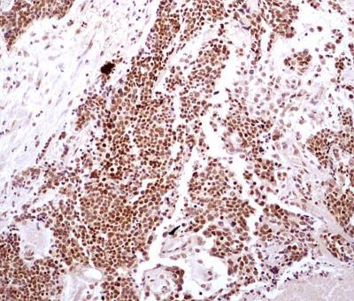
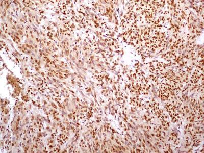
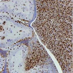
- Immunohistochemistry-Paraffin: HMGB1/HMG-1 Antibody (19N10B7) [NBP2-27396] - IHC-P detection of HMGB1 protein in a formalin-fixed paraffin-embedded tissue section of human bladder cancer using HMGB1 antibody (clone 19N10B7) at 5 ug/ml concentration. Strong nuclear HMGB1 immunopositivity was observed in the bladder cancer cells whereas the staining was weak in cells of tumor stroma.
- Submitted by
- Novus Biologicals (provider)
- Main image
- Experimental details
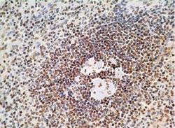
- Immunohistochemistry-Paraffin: HMGB1/HMG-1 Antibody (19N10B7) [NBP2-27396] - Formalin-fixed, paraffin-embedded human spleen tissue stained with HMGB1 antibody (5 ug/ml), peroxidase-conjugate and DAB chromogen. Staining of formalin-fixed tissues is enhanced by boiling tissue sections in 10 mM sodium citrate buffer, pH 6.0 for 10-20 min followed by cooling at RT for 20 min.

- Submitted by
- Novus Biologicals (provider)
- Main image
- Experimental details
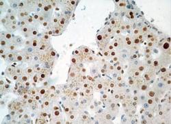
- Immunohistochemistry-Paraffin: HMGB1/HMG-1 Antibody (19N10B7) [NBP2-27396] - Formalin-fixed, paraffin-embedded human liver tissue stained with HMGB1 antibody (5 ug/ml), peroxidase-conjugate and DAB chromogen. Staining of formalin-fixed tissues is enhanced by boiling tissue sections in 10 mM sodium citrate buffer, pH 6.0 for 10-20 min followed by cooling at RT for 20 min.

- Submitted by
- Novus Biologicals (provider)
- Main image
- Experimental details
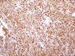
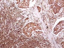
- Immunohistochemistry-Paraffin: HMGB1/HMG-1 Antibody (19N10B7) [NBP2-27396] - IHC-P detection of HMGB1 protein in a formalin-fixed paraffin-embedded tissue section of malignant stromal tumor of small bowel from human using HMGB1 antibody (clone 19N10B7) at 5 ug/ml concentration. HMGB1 immunopositivity of differential intensity was observed in the cells of tested section.
- Submitted by
- Novus Biologicals (provider)
- Main image
- Experimental details
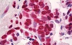
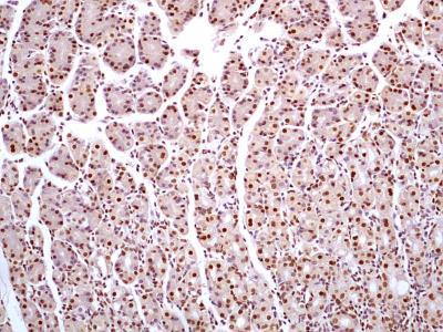
- Immunohistochemistry-Paraffin: HMGB1/HMG-1 Antibody (19N10B7) [NBP2-27396] - IHC-P detection of HMGB1 protein in a formalin-fixed paraffin-embedded tissue section of human stomach cancer using HMGB1 antibody (clone 19N10B7) at 5 ug/ml concentration. This representative image shows a distinct nuclear HMGB1 immunopositivity in the cancerous and sub-mucosal cells.

- Submitted by
- Novus Biologicals (provider)
- Main image
- Experimental details
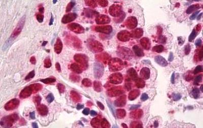
- Immunohistochemistry-Paraffin: HMGB1/HMG-1 Antibody (19N10B7) [NBP2-27396] - Formalin-fixed paraffin-embedded human prostate tissue stained with HMGB1 antibody at 10 ug/ml concentration. Staining of formalin-fixed tissues is enhanced by boiling tissue sections in 10 mM sodium citrate buffer, pH 6.0 for 10-20 min followed by cooling at RT for 20 min.
- Submitted by
- Novus Biologicals (provider)
- Main image
- Experimental details
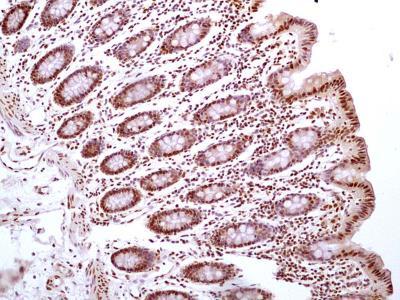
- Immunohistochemistry-Paraffin: HMGB1/HMG-1 Antibody (19N10B7) [NBP2-27396] - IHC-P detection of HMGB1 protein in a formalin-fixed paraffin-embedded tissue section of normal human colon using HMGB1 antibody (clone 19N10B7) at 5 ug/ml concentration. Representative image shows specific HMGB1 nuclear positivity in different mucosal cells of the colon.
- Submitted by
- Novus Biologicals (provider)
- Main image
- Experimental details
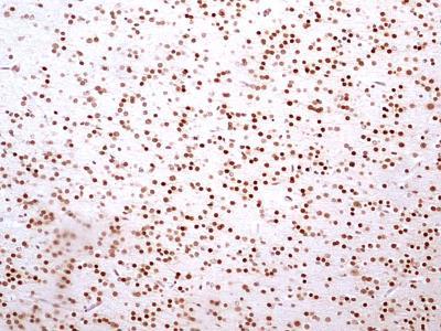
- Immunohistochemistry-Paraffin: HMGB1/HMG-1 Antibody (19N10B7) [NBP2-27396] - IHC-P detection of HMGB1 protein in a formalin-fixed paraffin-embedded tissue section of normal human brain using HMGB1 antibody (clone 19N10B7) at 5 ug/ml concentration. Representative image shows a distinct nuclear immunostaining of HMGB1 in the various brain cells.
- Submitted by
- Novus Biologicals (provider)
- Main image
- Experimental details
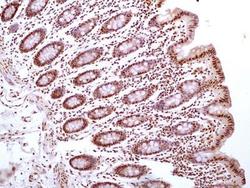
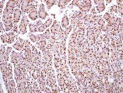
- Immunohistochemistry-Paraffin: HMGB1/HMG-1 Antibody (19N10B7) [NBP2-27396] - IHC-P detection of HMGB1 protein in a formalin-fixed paraffin-embedded tissue section of human stomach using HMGB1 antibody (clone 19N10B7) at 5 ug/ml concentration. Distinct nuclear staining of HMGB1 was observed in different cells of the glandular stomach.
- Submitted by
- Novus Biologicals (provider)
- Main image
- Experimental details
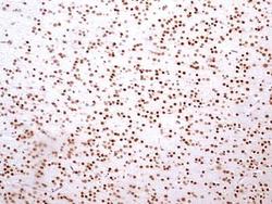
- Immunohistochemistry-Paraffin: HMGB1/HMG-1 Antibody (19N10B7) [NBP2-27396] - Analysis of formalin-fixed, decalcified, paraffin embedded tissue section from the paws of mouse (collagen-induced arthritis model) using HMGB1 antibody (clone 19N10B7) at 5 ug/ml concentration. [Image courtesy of Dr Ulf Andersson, Karolinska Institute].

- Submitted by
- Novus Biologicals (provider)
- Main image
- Experimental details
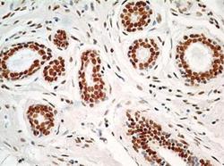
- Immunohistochemistry-Paraffin: HMGB1/HMG-1 Antibody (19N10B7) [NBP2-27396] - Formalin-fixed, paraffin-embedded human breast stained with HMGB1 antibody at 5 ug/ml.

Supportive validation
- Submitted by
- Novus Biologicals (provider)
- Main image
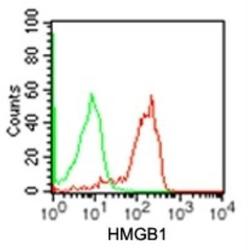
- Experimental details
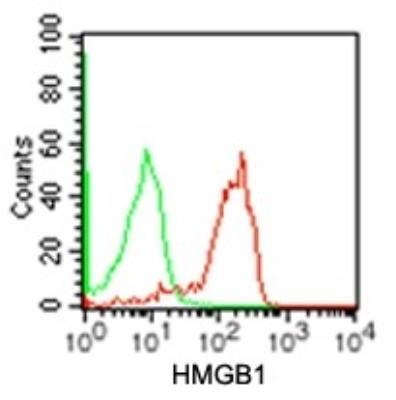
- Flow Cytometry: HMGB1/HMG-1 Antibody (19N10B7) [NBP2-27396] - Intracellular analysis using HMGB1 antibody. Human Jurkat cells were probed using 0.5 ug of HMGB1 antibody (red) and 0.5 ug of isotype control antibody (green).