Explore
 Validate
 Learn
Learn Western blot
 Immunocytochemistry
Antibody data
- Antibody Data
- Antigen structure
- References [13]
- Comments [0]
- Validations
- Western blot [9]
- Immunocytochemistry [4]
- Immunohistochemistry [4]
Submit
Validation data
Reference
Comment
Report error
- Product number
- GTX101277 - Provider product page

- Provider
- GeneTex
- Proper citation
- GeneTex Cat#GTX101277, RRID:AB_1950499
- Product name
- HMGB1 antibody
- Antibody type
- Polyclonal
- Reactivity
- Human, Mouse, Rat, Porcine
- Host
- Rabbit
Submitted references Heparanase regulates the M1 polarization of renal macrophages and their crosstalk with renal epithelial tubular cells after ischemia/reperfusion injury.
HMGB1/IL-1β complexes regulate neuroimmune responses in alcoholism.
MicroRNA-200c inhibits epithelial-mesenchymal transition, invasion, and migration of lung cancer by targeting HMGB1.
Dual therapeutic functions of F-5 fragment in burn wounds: preventing wound progression and promoting wound healing in pigs.
Simultaneous induction of apoptosis and necroptosis by Tanshinone IIA in human hepatocellular carcinoma HepG2 cells.
Resources for the Comprehensive Discovery of Functional RNA Elements.
ASC filament formation serves as a signal amplification mechanism for inflammasomes.
Receptor for Advanced Glycation End Products and its Inflammatory Ligands are Upregulated in Amyotrophic Lateral Sclerosis.
Amitriptyline pharmacologically preconditions rat hearts against cardiac ischemic-reperfusion injury.
Shikonin-enhanced cell immunogenicity of tumor vaccine is mediated by the differential effects of DAMP components.
Caspase-11 activates a canonical NLRP3 inflammasome by promoting K(+) efflux.
High-mobility group box 1 expression and lymph node metastasis in intrahepatic cholangiocarcinoma.
Non-canonical inflammasome activation targets caspase-11.
Masola V, Zaza G, Bellin G, Dall'Olmo L, Granata S, Vischini G, Secchi MF, Lupo A, Gambaro G, Onisto M
FASEB journal : official publication of the Federation of American Societies for Experimental Biology 2018 Feb;32(2):742-756
FASEB journal : official publication of the Federation of American Societies for Experimental Biology 2018 Feb;32(2):742-756
HMGB1/IL-1β complexes regulate neuroimmune responses in alcoholism.
Coleman LG Jr, Zou J, Qin L, Crews FT
Brain, behavior, and immunity 2018 Aug;72:61-77
Brain, behavior, and immunity 2018 Aug;72:61-77
MicroRNA-200c inhibits epithelial-mesenchymal transition, invasion, and migration of lung cancer by targeting HMGB1.
Liu PL, Liu WL, Chang JM, Chen YH, Liu YP, Kuo HF, Hsieh CC, Ding YS, Chen WW, Chong IW
PloS one 2017;12(7):e0180844
PloS one 2017;12(7):e0180844
Dual therapeutic functions of F-5 fragment in burn wounds: preventing wound progression and promoting wound healing in pigs.
Bhatia A, O'Brien K, Chen M, Wong A, Garner W, Woodley DT, Li W
Molecular therapy. Methods & clinical development 2016;3:16041
Molecular therapy. Methods & clinical development 2016;3:16041
Simultaneous induction of apoptosis and necroptosis by Tanshinone IIA in human hepatocellular carcinoma HepG2 cells.
Lin CY, Chang TW, Hsieh WH, Hung MC, Lin IH, Lai SC, Tzeng YJ
Cell death discovery 2016;2:16065
Cell death discovery 2016;2:16065
Resources for the Comprehensive Discovery of Functional RNA Elements.
Sundararaman B, Zhan L, Blue SM, Stanton R, Elkins K, Olson S, Wei X, Van Nostrand EL, Pratt GA, Huelga SC, Smalec BM, Wang X, Hong EL, Davidson JM, Lécuyer E, Graveley BR, Yeo GW
Molecular cell 2016 Mar 17;61(6):903-13
Molecular cell 2016 Mar 17;61(6):903-13
ASC filament formation serves as a signal amplification mechanism for inflammasomes.
Dick MS, Sborgi L, Rühl S, Hiller S, Broz P
Nature communications 2016 Jun 22;7:11929
Nature communications 2016 Jun 22;7:11929
Receptor for Advanced Glycation End Products and its Inflammatory Ligands are Upregulated in Amyotrophic Lateral Sclerosis.
Juranek JK, Daffu GK, Wojtkiewicz J, Lacomis D, Kofler J, Schmidt AM
Frontiers in cellular neuroscience 2015;9:485
Frontiers in cellular neuroscience 2015;9:485
Amitriptyline pharmacologically preconditions rat hearts against cardiac ischemic-reperfusion injury.
Lee SM, Hutchinson M, Staikopoulos V, Saint DA
International journal of cardiology 2015;190:353-9
International journal of cardiology 2015;190:353-9
Shikonin-enhanced cell immunogenicity of tumor vaccine is mediated by the differential effects of DAMP components.
Lin TJ, Lin HT, Chang WT, Mitapalli S P, Hsiao PW, Yin SY, Yang NS
Molecular cancer 2015 Sep 24;14:174
Molecular cancer 2015 Sep 24;14:174
Caspase-11 activates a canonical NLRP3 inflammasome by promoting K(+) efflux.
Rühl S, Broz P
European journal of immunology 2015 Oct;45(10):2927-36
European journal of immunology 2015 Oct;45(10):2927-36
High-mobility group box 1 expression and lymph node metastasis in intrahepatic cholangiocarcinoma.
Xu YF, Ge FJ, Han B, Yang XQ, Su H, Zhao AC, Zhao MH, Yang YB, Yang J
World journal of gastroenterology 2015 Mar 21;21(11):3256-65
World journal of gastroenterology 2015 Mar 21;21(11):3256-65
Non-canonical inflammasome activation targets caspase-11.
Kayagaki N, Warming S, Lamkanfi M, Vande Walle L, Louie S, Dong J, Newton K, Qu Y, Liu J, Heldens S, Zhang J, Lee WP, Roose-Girma M, Dixit VM
Nature 2011 Oct 16;479(7371):117-21
Nature 2011 Oct 16;479(7371):117-21
No comments: Submit comment
Enhanced validation
Supportive validation
- Submitted by
- GeneTex (provider)
- Enhanced method
- Genetic validation
- Main image
- Experimental details
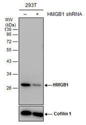
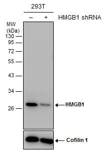
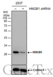
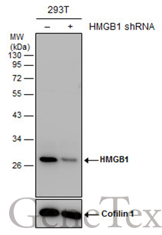
- Non-transfected (¡V) and transfected (+) 293T whole cell extracts (30 ?g) were separated by 12% SDS-PAGE, and the membrane was blotted with HMGB1 antibody (GTX101277) diluted at 1:5000. The HRP-conjugated anti-rabbit IgG antibody (GTX213110-01) was used to detect the primary antibody.
Supportive validation
- Submitted by
- GeneTex (provider)
- Main image
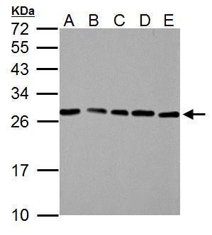
- Experimental details
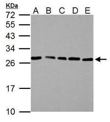
- HMGB1 antibody detects HMGB1 protein by western blot analysis.A. 30 ?g NIH-3T3 whole cell lysate/extractB. 30 ?g JC whole cell lysate/extractC. 30 ?g BCL-1 whole cell lysate/extractD. 30 ?g C2C12 whole cell lysate/extractE. 30 ?g Raw264.7 whole cell lysate/extract12% SDS-PAGEHMGB1 antibody (GTX101277) dilution: 1:3000 The HRP-conjugated anti-rabbit IgG antibody (GTX213110-01) was used to detect the primary antibody.
- Submitted by
- GeneTex (provider)
- Main image
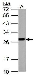
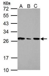
- Experimental details
- HMGB1 antibody detects HMGB1 protein by western blot analysis.A. 30 ?g PC-12 whole cell lysate/extract12% SDS-PAGEHMGB1 antibody (GTX101277) dilution: 1:3000 The HRP-conjugated anti-rabbit IgG antibody (GTX213110-01) was used to detect the primary antibody.
- Submitted by
- GeneTex (provider)
- Main image
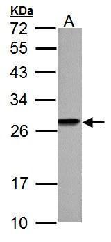
- Experimental details
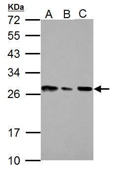
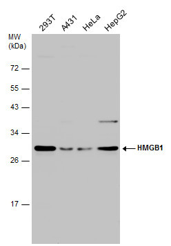
- HMGB1 antibody detects HMGB1 protein by western blot analysis.A. 30 ?g 293T whole cell lysate/extract B. 30 ?g A431 whole cell lysate/extractC. 30 ?g A375 whole cell lysate/extract12% SDS-PAGEHMGB1 antibody (GTX101277) dilution: 1:3000 The HRP-conjugated anti-rabbit IgG antibody (GTX213110-01) was used to detect the primary antibody.
- Submitted by
- GeneTex (provider)
- Main image
- Experimental details
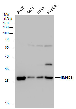
- HMGB1 antibody detects HMGB1 protein by western blot analysis. Various whole cell extracts (30 ?g) were separated by 12% SDS-PAGE, and the membrane was blotted with HMGB1 antibody (GTX101277) diluted by 1:3000.
- Validation comment
- WB
- Submitted by
- GeneTex (provider)
- Main image
- Experimental details
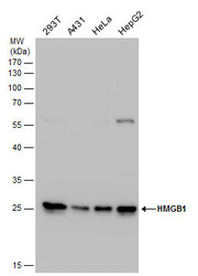
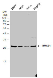
- HMGB1 antibody detects HMGB1 protein by western blot analysis. Various whole cell extracts (30 ?g) were separated by 12% SDS-PAGE, and the membrane was blotted with HMGB1 antibody (GTX101277) diluted by 1:3000. The HRP-conjugated anti-rabbit IgG antibody (GTX213110-01) was used to detect the primary antibody.
- Submitted by
- GeneTex (provider)
- Main image
- Experimental details
- Non-transfected (¡V) and transfected (+) 293T whole cell extracts (30 ?g) were separated by 12% SDS-PAGE, and the membrane was blotted with HMGB1 antibody (GTX101277) diluted at 1:5000. The HRP-conjugated anti-rabbit IgG antibody (GTX213110-01) was used to detect the primary antibody.
- Submitted by
- GeneTex (provider)
- Main image
- Experimental details
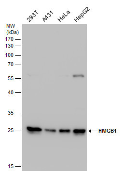
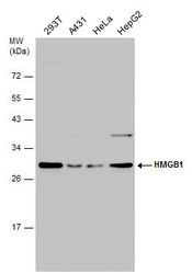
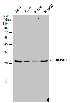
- Various whole cell extracts (30 ?g) were separated by 12% SDS-PAGE, and the membrane was blotted with HMGB1 antibody (GTX101277) diluted at 1:3000.
- Submitted by
- GeneTex (provider)
- Main image
- Experimental details
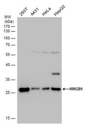
- Various whole cell extracts (30 ?g) were separated by 12% SDS-PAGE, and the membrane was blotted with HMGB1 antibody (GTX101277) diluted at 1:3000.
Supportive validation
- Submitted by
- GeneTex (provider)
- Main image
- Experimental details
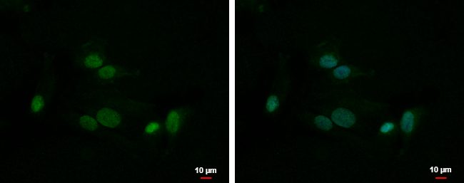
- HMGB1 antibody detects HMGB1 protein at nucleus by immunofluorescent analysis.Sample: SK-N-SH cells were fixed in 4% paraformaldehyde at RT for 15 min.Green: HMGB1 protein stained by HMGB1 antibody (GTX101277) diluted at 1:500.Blue: Hoechst 33342 staining.Scale bar = 10 £gm.
- Submitted by
- GeneTex (provider)
- Main image
- Experimental details
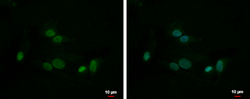
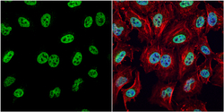
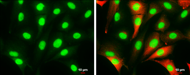
- HMGB1 antibody detects HMGB1 protein at nucleus by immunofluorescent analysis.Sample: HeLa cells were fixed in 4% paraformaldehyde at RT for 15 min.Green: HMGB1 protein stained by HMGB1 antibody (GTX101277) diluted at 1:1000.Red: phalloidin, a cytoskeleton marker, stained by phalloidin (invitrogen, A12380) diluted at 1:200.Blue: Hoechst 33342 staining.

- Submitted by
- GeneTex (provider)
- Main image
- Experimental details
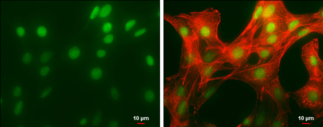
- HMGB1 antibody detects HMGB1 protein at nucleus by immunofluorescent analysis.Sample: NIH/3T3 cells were fixed in 4% paraformaldehyde at RT for 15 min.Green: HMGB1 protein stained by HMGB1 antibody (GTX101277) diluted at 1:500.Red: phalloidin, a cytoskeleton marker, diluted at 1:50.Scale bar = 10 £gm.
- Submitted by
- GeneTex (provider)
- Main image

- Experimental details
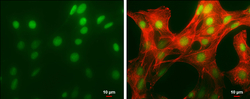
- HMGB1 antibody detects HMGB1 protein at cytoplasm and nucleus by immunofluorescent analysis.Sample: SK-N-SH cells were fixed in 4% paraformaldehyde at RT for 15 min.Green: HMGB1 protein stained by HMGB1 antibody (GTX101277) diluted at 1:1000.Red: beta Tubulin 3/ Tuj1, a cytoskeleton marker, stained by beta Tubulin 3/ Tuj1 antibody [GT11710] (GTX631836) diluted at 1:500.Scale bar = 10 £gm.
Supportive validation
- Submitted by
- GeneTex (provider)
- Main image

- Experimental details
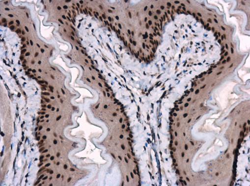
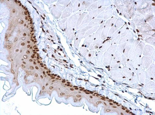
- HMGB1 antibody detects HMGB1 protein at nucleus on mouse esophagus by immunohistochemical analysis. Sample: Paraffin-embedded mouse esophagus. HMGB1 antibody (GTX101277) dilution: 1:1000.
- Submitted by
- GeneTex (provider)
- Main image
- Experimental details
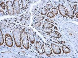
- HMGB1 antibody detects HMGB1 protein at nucleus on mouse colon by immunohistochemical analysis. Sample: Paraffin-embedded mouse colon. HMGB1 antibody (GTX101277) dilution: 1:1000.

- Submitted by
- GeneTex (provider)
- Main image
- Experimental details
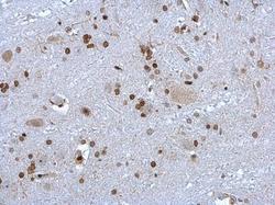
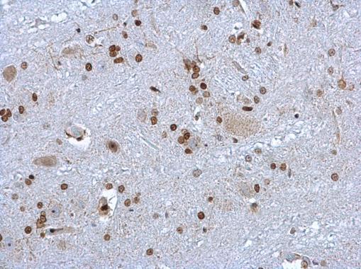
- HMGB1 antibody detects HMGB1 protein at nucleus on rat brain stem by immunohistochemical analysis. Sample: Paraffin-embedded rat brain stem. HMGB1 antibody (GTX101277) dilution: 1:1000.
- Submitted by
- GeneTex (provider)
- Main image

- Experimental details
- HMGB1 antibody detects HMGB1 protein at nucleus in mouse esophagus by immunohistochemical analysis. Sample: Paraffin-embedded mouse esophagus. HMGB1 antibody (GTX101277) diluted at 1:1000.