Explore
 Validate
 Learn
Learn Western blot
 Immunocytochemistry
Antibody data
- Antibody Data
- Antigen structure
- References [1]
- Comments [0]
- Validations
- Western blot [6]
- Immunocytochemistry [1]
- Immunohistochemistry [3]
Submit
Validation data
Reference
Comment
Report error
- Product number
- GTX112959 - Provider product page

- Provider
- GeneTex
- Proper citation
- GeneTex Cat#GTX112959, RRID:AB_1950500
- Product name
- HMGB1 antibody
- Antibody type
- Polyclonal
- Reactivity
- Human, Mouse, Rat
- Host
- Rabbit
Submitted references Tumor-derived exosomes confer antigen-specific immunosuppression in a murine delayed-type hypersensitivity model.
Yang C, Kim SH, Bianco NR, Robbins PD
PloS one 2011;6(8):e22517
PloS one 2011;6(8):e22517
No comments: Submit comment
Enhanced validation
Supportive validation
- Submitted by
- GeneTex (provider)
- Enhanced method
- Genetic validation
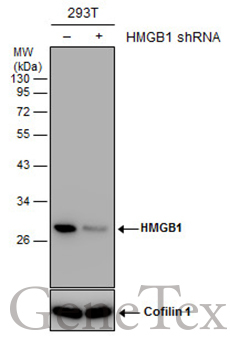
- Main image
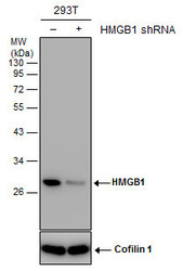
- Experimental details
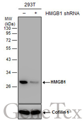
- Non-transfected (¡V) and transfected (+) 293T whole cell extracts (30 ?g) were separated by 12% SDS-PAGE, and the membrane was blotted with HMGB1 antibody (GTX112959) diluted at 1:2000. The HRP-conjugated anti-rabbit IgG antibody (GTX213110-01) was used to detect the primary antibody.
Supportive validation
- Submitted by
- GeneTex (provider)
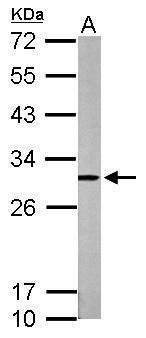
- Main image

- Experimental details
- Sample (50 ?g of whole cell lysate) A: mouse brain 12% SDS PAGE GTX112959 diluted at 1:1000 The HRP-conjugated anti-rabbit IgG antibody (GTX213110-01) was used to detect the primary antibody.
- Submitted by
- GeneTex (provider)
- Main image

- Experimental details
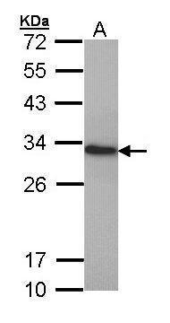
- Sample (30 ?g of whole cell lysate) A: HepG2 (GTX27900) 12% SDS PAGE GTX112959 diluted at 1:1000 The HRP-conjugated anti-rabbit IgG antibody (GTX213110-01) was used to detect the primary antibody.
- Submitted by
- GeneTex (provider)
- Main image

- Experimental details
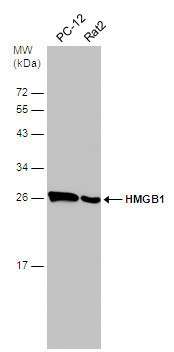
- Various whole cell extracts (30 ?g) were separated by 12% SDS-PAGE, and the membrane was blotted with HMGB1 antibody (GTX112959) diluted at 1:500. The HRP-conjugated anti-rabbit IgG antibody (GTX213110-01) was used to detect the primary antibody.
- Submitted by
- GeneTex (provider)
- Main image
- Experimental details
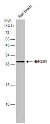
- Rat tissue extract (50 ?g) was separated by 12% SDS-PAGE, and the membrane was blotted with HMGB1 antibody (GTX112959) diluted at 1:500. The HRP-conjugated anti-rabbit IgG antibody (GTX213110-01) was used to detect the primary antibody.

- Submitted by
- GeneTex (provider)
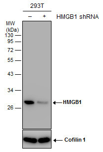
- Main image
- Experimental details
- Non-transfected (¡V) and transfected (+) 293T whole cell extracts (30 ?g) were separated by 12% SDS-PAGE, and the membrane was blotted with HMGB1 antibody (GTX112959) diluted at 1:2000. The HRP-conjugated anti-rabbit IgG antibody (GTX213110-01) was used to detect the primary antibody.
Supportive validation
- Submitted by
- GeneTex (provider)
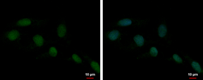
- Main image

- Experimental details
- HMGB1 antibody detects HMGB1 protein at nucleus by immunofluorescent analysis.Sample: NT2D1 cells were fixed in 4% paraformaldehyde at RT for 15 min.Green: HMGB1 protein stained by HMGB1 antibody (GTX112959) diluted at 1:1000.Blue: Hoechst 33342 staining.
Supportive validation
- Submitted by
- GeneTex (provider)
- Main image
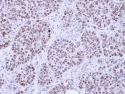
- Experimental details
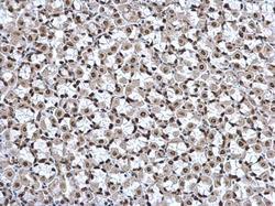
- Immunohistochemical analysis of paraffin-embedded SW480 Xenograft, using HMG-1(GTX112959) antibody at 1:100 dilution.
- Submitted by
- GeneTex (provider)
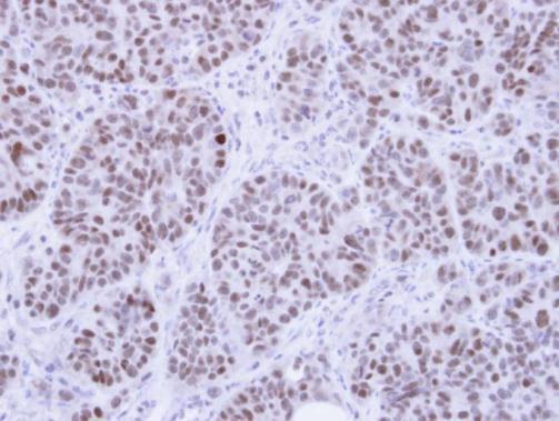
- Main image
- Experimental details
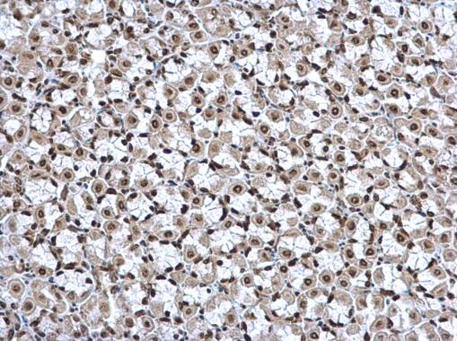
- HMGB1 antibody detects HMGB1 protein at nucleus on mouse stomach by immunohistochemical analysis. Sample: Paraffin-embedded mouse stomach. HMGB1 antibody (GTX112959) dilution: 1:500.
- Submitted by
- GeneTex (provider)
- Main image
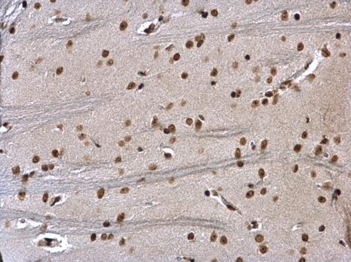
- Experimental details
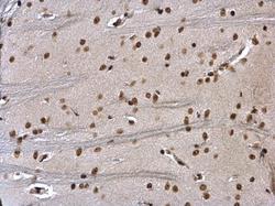
- HMGB1 antibody detects HMGB1 protein at nucleus on rat fore brain by immunohistochemical analysis. Sample: Paraffin-embedded rat fore brain. HMGB1 antibody (GTX112959) dilution: 1:500.