Explore
 Validate
 Learn
Learn Western blot
Antibody data
- Antibody Data
- Antigen structure
- References [4]
- Comments [0]
- Validations
- Western blot [7]
- Immunocytochemistry [2]
- Immunohistochemistry [6]
- Other assay [7]
Submit
Validation data
Reference
Comment
Report error
- Product number
- MA5-17278 - Provider product page

- Provider
- Invitrogen Antibodies
- Product name
- HMGB1 Monoclonal Antibody (GT348)
- Antibody type
- Monoclonal
- Antigen
- Recombinant protein fragment
- Description
- Recommended positive controls: 293T, A431, HepG2, Neuro2A, C8D30, NIH-3T3, BCL-1, Raw264.7, C2C12, PC-12, Rat2, mouse brain, rat brain. Predicted reactivity: Mouse (100%), Rat (100%), Xenopus laevis (93%), Dog (100%), Pig (100%), Rabbit (100%), Chicken (94%), Rhesus Monkey (100%), Bovine (100%). Store product as a concentrated solution. Centrifuge briefly prior to opening the vial.
- Reactivity
- Human, Mouse, Rat
- Host
- Mouse
- Isotype
- IgG
- Antibody clone number
- GT348
- Vial size
- 100 µL
- Concentration
- 0.88 mg/mL
- Storage
- Store at 4°C short term. For long term storage, store at -20°C, avoiding freeze/thaw cycles.
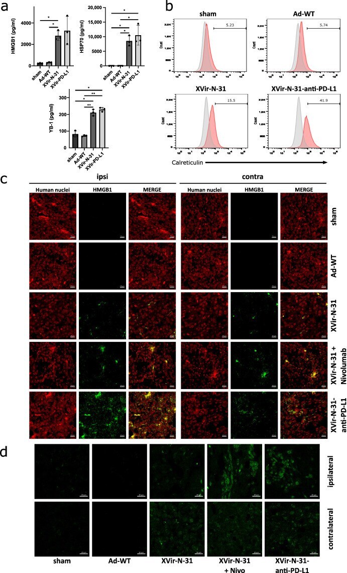
Submitted references The Oncolytic Adenovirus XVir-N-31, in Combination with the Blockade of the PD-1/PD-L1 Axis, Conveys Abscopal Effects in a Humanized Glioblastoma Mouse Model.
ABT‑737, a Bcl‑2 family inhibitor, has a synergistic effect with apoptosis by inducing urothelial carcinoma cell necroptosis.
Alpha-Synuclein Preformed Fibrils Induce Cellular Senescence in Parkinson's Disease Models.
Molecular insights on cytochrome c and nucleotide regulation of apoptosome function and its implication in cancer.
Klawitter M, El-Ayoubi A, Buch J, Rüttinger J, Ehrenfeld M, Lichtenegger E, Krüger MA, Mantwill K, Koll FJ, Kowarik MC, Holm PS, Naumann U
International journal of molecular sciences 2022 Sep 1;23(17)
International journal of molecular sciences 2022 Sep 1;23(17)
ABT‑737, a Bcl‑2 family inhibitor, has a synergistic effect with apoptosis by inducing urothelial carcinoma cell necroptosis.
Cheng R, Liu X, Wang Z, Tang K
Molecular medicine reports 2021 Jun;23(6)
Molecular medicine reports 2021 Jun;23(6)
Alpha-Synuclein Preformed Fibrils Induce Cellular Senescence in Parkinson's Disease Models.
Verma DK, Seo BA, Ghosh A, Ma SX, Hernandez-Quijada K, Andersen JK, Ko HS, Kim YH
Cells 2021 Jul 5;10(7)
Cells 2021 Jul 5;10(7)
Molecular insights on cytochrome c and nucleotide regulation of apoptosome function and its implication in cancer.
Yadav N, Gogada R, O'Malley J, Gundampati RK, Jayanthi S, Hashmi S, Lella R, Zhang D, Wang J, Kumar R, Suresh Kumar TK, Chandra D
Biochimica et biophysica acta. Molecular cell research 2020 Jan;1867(1):118573
Biochimica et biophysica acta. Molecular cell research 2020 Jan;1867(1):118573
No comments: Submit comment
Supportive validation
- Submitted by
- Invitrogen Antibodies (provider)
- Main image
- Experimental details
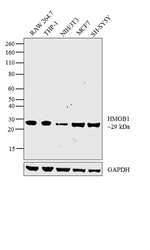
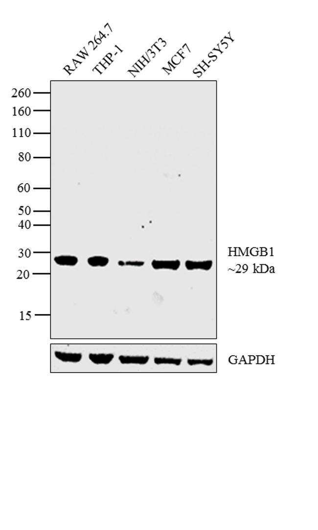
- Western blot analysis was performed on whole cell extracts (30 µg lysate) of RAW 264.7 (Lane 1), THP-1 (Lane 2), NIH/3T3 (Lane 3), MCF7 (Lane 4) and SH-SY5Y (Lane 5). The blot was probed with Anti-HMGB1 Monoclonal Antibody (Product # MA5-17278, 1 µg/ml) and detected by chemiluminescence using Goat anti-Mouse IgG (H+L) Superclonal™ Secondary Antibody, HRP conjugate (Product # A28177, 0.25 µg/ml, 1:4000 dilution). A 29 kDa band corresponding to HMGB1 was observed across the cell lines tested.
- Submitted by
- Invitrogen Antibodies (provider)
- Main image
- Experimental details
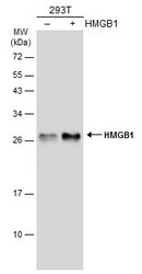
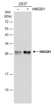
- Western Blot analysis of HMGB1 was performed by separating 30 µg of non-transfected (–) and transfected (+) 293T whole cell extracts by 12% SDS-PAGE. Proteins were transferred to a membrane and probed with a HMGB1 Monoclonal Antibody (GT348) (Product # MA5-17278) at a dilution of 1:3000. The HRP-conjugated anti-mouset IgG antibody was used to detect the primary antibody.
- Submitted by
- Invitrogen Antibodies (provider)
- Main image

- Experimental details
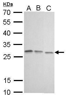
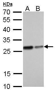
- HMGB1 Monoclonal Antibody (GT348) detects HMGB1 protein by western blot analysis. A. 30 µg 293T whole cell lysate/extract. B. 30 µg A431 whole cell lysate/extract. C. 30 µg HepG2 whole cell lysate/extract.12% SDS-PAGE. HMGB1 Monoclonal Antibody (GT348) (Product # MA5-17278) dilution: 1:1,000. The HRP-conjugated anti-mouse IgG antibody was used to detect the primary antibody.
- Submitted by
- Invitrogen Antibodies (provider)
- Main image
- Experimental details
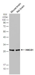
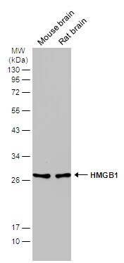
- Western blot analysis of HMGB1 was performed by separating 50 µg of various tissue extracts by 12% SDS-PAGE. Proteins were transferred to a membrane and probed with a HMGB1 Monoclonal Antibody (GT348) (Product # MA5-17278) at a dilution of 1:2000. The HRP-conjugated anti-mouse IgG antibody was used to detect the primary antibody.
- Submitted by
- Invitrogen Antibodies (provider)
- Main image

- Experimental details
- HMGB1 Monoclonal Antibody (GT348) detects HMGB1 protein by western blot analysis. A. 30 µg Neuro2A whole cell lysate/extract. B. 30 µg C8D30 whole cell lysate/extract. C. 30 µg NIH-3T3 whole cell lysate/extract. D. 30 µg BCL-1 whole cell lysate/extract. E. 30 µg Raw264.7 whole cell lysate/extract. F. 30 µg C2C12 whole cell lysate/extract.12% SDS-PAGE. HMGB1 Monoclonal Antibody (GT348) (Product # MA5-17278) dilution: 1:500. The HRP-conjugated anti-mouse IgG antibody was used to detect the primary antibody.

- Submitted by
- Invitrogen Antibodies (provider)
- Main image

- Experimental details
- HMGB1 Monoclonal Antibody (GT348) detects HMGB1 protein by western blot analysis. A. 30 µg PC-12 whole cell lysate/extract. B. 30 µg Rat-2 whole cell lysate/extract.12% SDS-PAGE. HMGB1 Monoclonal Antibody (GT348) (Product # MA5-17278) dilution: 1:1,000. The HRP-conjugated anti-mouse IgG antibody was used to detect the primary antibody.
- Submitted by
- Invitrogen Antibodies (provider)
- Main image
- Experimental details
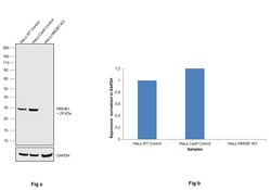
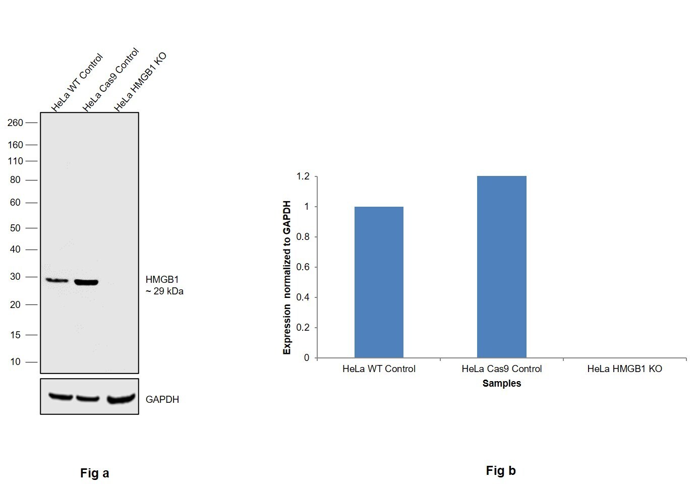
- Knockout of HMGB1 was achieved by CRISPR-Cas9 genome editing using LentiArray™ Lentiviral sgRNA (Product # A32042, Assay ID CRISPR784665_LV) and LentiArray Cas9 Lentivirus (Product # A32064). Western blot analysis of HMGB1 was performed by loading 30 µg of HeLa Wild Type (Lane 1), HeLa Cas9 (Lane 2) andHeLa HMGB1 KO (Lane 3) whole cell extracts. The samples were electrophoresed using NuPAGE™ Novex™ 4-12% Bis-Tris Protein Gel (Product # NP0322BOX). Resolved proteins were then transferred onto a nitrocellulose membrane (Product # IB23001) by iBlot® 2 Dry Blotting System (Product # IB21001). The blot was probed with Anti-HMGB1 Monoclonal Antibody (GT348) (Product # MA5-17278, 1 µg/mL dilution) and Goat anti-Mouse IgG (H+L) Superclonal™ Recombinant Secondary Antibody, HRP (Product # A28177, 1:5,000 dilution) using the iBright FL 1000 (Product # A32752). Chemiluminescent detection was performed using SuperSignal™ West Dura Extended Duration Substrate (Product # 34076). Loss of signal upon CRISPR mediated knockout (KO) using the LentiArray™ CRISPR product line confirms that antibody is specific to HMGB1.
Supportive validation
- Submitted by
- Invitrogen Antibodies (provider)
- Main image
- Experimental details
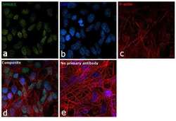
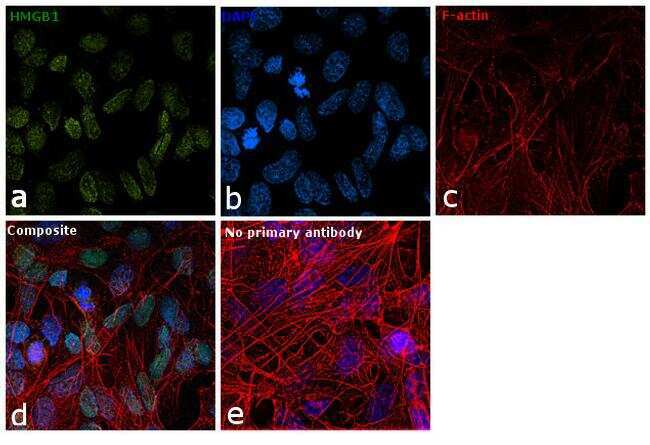
- Immunofluorescence analysis of HMGB1 was performed using 90% confluent log phase SH-SY5Y cells. The cells were fixed with 4% paraformaldehyde for 10 minutes, permeabilized with 0.1% Triton™ X-100 for 15 minutes, and blocked with 1% BSA for 1 hour at room temperature. The cells were labeled with HMGB1 Monoclonal Antibody (GT348) (Product # MA5-17278) at 5 µg/mL in 0.1% BSA, incubated at 4 degree Celsius overnight and then labeled with Goat anti-Mouse IgG (H+L) Superclonal™ Secondary Antibody, Alexa Fluor® 488 conjugate (Product # A28175) at a dilution of 1:2000 for 45 minutes at room temperature (Panel a: green). Nuclei (Panel b: blue) were stained with SlowFade® Gold Antifade Mountant with DAPI (Product # S36938). F-actin (Panel c: red) was stained with Rhodamine Phalloidin (Product # R415, 1:300). Panel d represents the merged image showing nuclear localization. Panel e represents control cells with no primary antibody to assess background. The images were captured at 60X magnification.
- Submitted by
- Invitrogen Antibodies (provider)
- Main image

- Experimental details
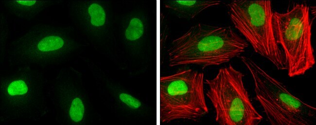
- Immunocytochemistry-Immunofluorescence analysis of HMGB1 was performed in HeLa cells fixed in 4% paraformaldehyde at RT for 15 min. Green: HMGB1 Monoclonal Antibody (GT348) (Product # MA5-17278) diluted at 1:200. Red: phalloidin, a cytoskeleton marker.
Supportive validation
- Submitted by
- Invitrogen Antibodies (provider)
- Main image

- Experimental details
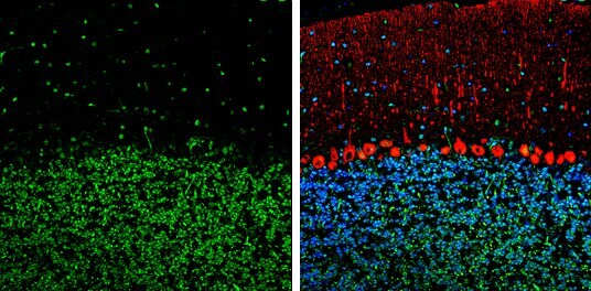
- Immunohistochemistry (Frozen) analysis of HMGB1 was performed in frozen-sectioned adult mouse cerebellum tissue using HMGB1 Monoclonal Antibody (GT348) (Product # MA5-17278) at a dilution of 1:250 (Green). Red: Calbindin, stained by Calbindin antibody diluted at 1:500. Blue: Fluoroshield with DAPI.
- Submitted by
- Invitrogen Antibodies (provider)
- Main image
- Experimental details
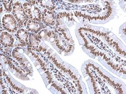
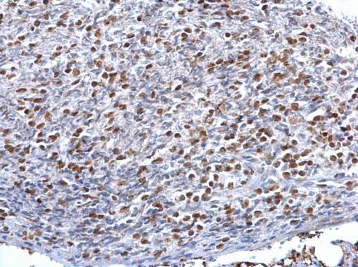
- HMGB1 Monoclonal Antibody (GT348) detects HMGB1 protein at nucleus on mouse lymph node by immunohistochemical analysis. Sample: Paraffin-embedded mouse lymph node. HMGB1 Monoclonal Antibody (GT348) (Product # MA5-17278) dilution: 1:500. Antigen Retrieval: EDTA based buffer, pH 8.0, 15 min.
- Submitted by
- Invitrogen Antibodies (provider)
- Main image
- Experimental details
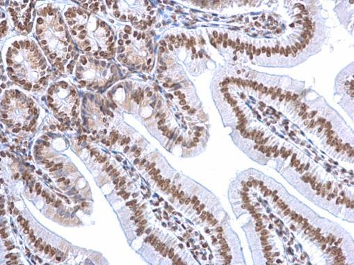
- Immunohistochemical analysis of HMGB1 was performed using formalin-fixed paraffin-embedded human colon adenocarcinoma tissue sections. To expose the target protein, heat-induced epitope retrieval was performed on de-paraffinized sections using eBioscience™ IHC Antigen Retrieval Solution - Low pH (10X) (Product # 00-4955-58) diluted to 1X solution in water in a decloaking chamber at 110 degree Celsius for 15 minutes. Following antigen retrieval, the sections were blocked with 2% normal goat serum in 1X PBS for 45 minutes at room temperature and then probed with or without HMGB1 Monoclonal Antibody (GT348) (Product # MA5-17278) at 1:100 dilution in 0.1% normal goat serum overnight at 4 degree Celsius in a humidified chamber. Detection was performed using Goat anti-Mouse IgG (H+L) Highly Cross-Adsorbed Secondary Antibody, Alexa Fluor Plus 488 (Product # A32723) at a dilution of 1:2000 in 0.1% normal goat serum for 45 minutes at room temperature. ReadyProbes™ Tissue Autofluorescence Quenching Kit (Product # R37630) was used to quench autofluorescence from the tissues. Nuclei were stained with DAPI (Product # D1306) and the sections were mounted using ProLong™ Glass Antifade Mountant (Product # P36984). The images were captured on EVOS™ M7000 Imaging System (Product # AMF7000) at 20X magnification.

- Submitted by
- Invitrogen Antibodies (provider)
- Main image

- Experimental details
- HMGB1 Monoclonal Antibody (GT348) detects HMGB1 protein at nucleus on mouse lymph node by immunohistochemical analysis. Sample: Paraffin-embedded mouse lymph node. HMGB1 Monoclonal Antibody (GT348) (Product # MA5-17278) dilution: 1:500. Antigen Retrieval: EDTA based buffer, pH 8.0, 15 min.
- Submitted by
- Invitrogen Antibodies (provider)
- Main image

- Experimental details
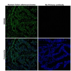
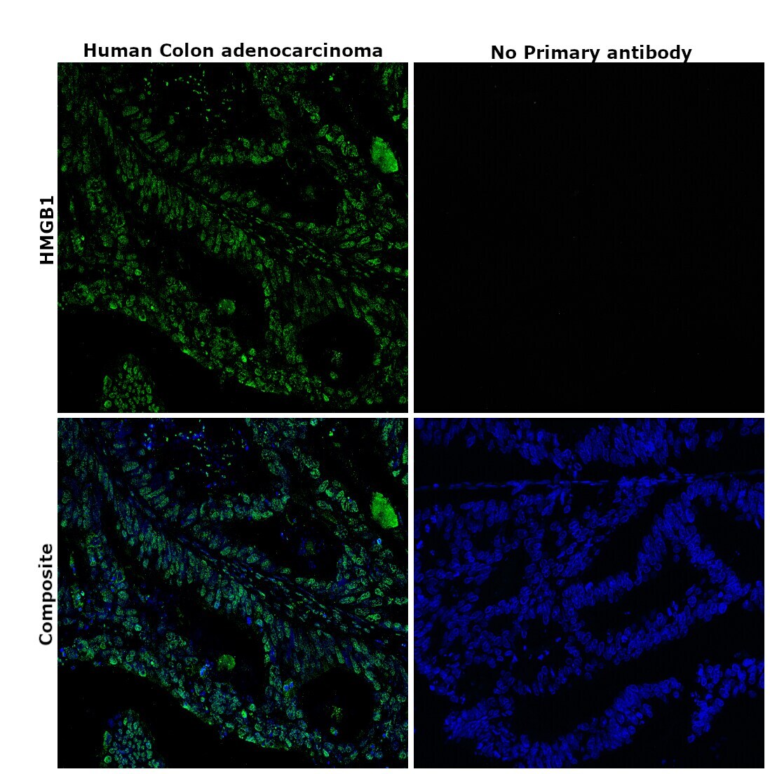
- Immunohistochemical analysis of HMGB1 was performed using formalin-fixed paraffin-embedded human colon adenocarcinoma tissue sections. To expose the target protein, heat-induced epitope retrieval was performed on de-paraffinized sections using eBioscience™ IHC Antigen Retrieval Solution - High pH (10X) (Product # 00-4956-58) diluted to 1X solution in water in a decloaking chamber at 110 degree Celsius for 15 minutes. Following antigen retrieval, the sections were blocked with 2% normal goat serum in 1X PBS for 45 minutes at room temperature and then probed with or without HMGB1 Monoclonal Antibody (GT348) (Product # MA5-17278) at 1:100 dilution in 0.1% normal goat serum overnight at 4 degree Celsius in a humidified chamber. Detection was performed using Goat anti-Mouse IgG (H+L) Highly Cross-Adsorbed Secondary Antibody, Alexa Fluor Plus 488 (Product # A32723) at a dilution of 1:2000 in 0.1% normal goat serum for 45 minutes at room temperature. ReadyProbes™ Tissue Autofluorescence Quenching Kit (Product # R37630) was used to quench autofluorescence from the tissues. Nuclei were stained with DAPI (Product # D1306) and the sections were mounted using ProLong™ Glass Antifade Mountant (Product # P36984). The images were captured on EVOS™ M7000 Imaging System (Product # AMF7000) at 20X magnification.
- Submitted by
- Invitrogen Antibodies (provider)
- Main image

- Experimental details
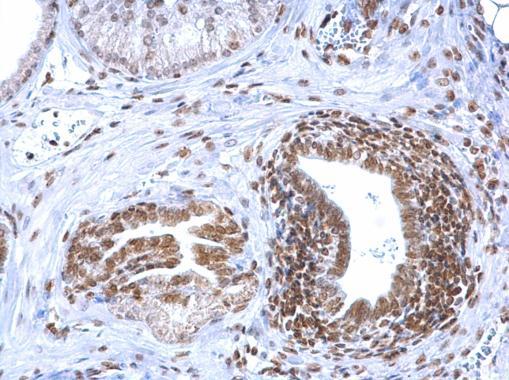
- HMGB1 Monoclonal Antibody (GT348) detects HMGB1 protein at nucleus on mouse prostate by immunohistochemical analysis. Sample: Paraffin-embedded mouse prostate. HMGB1 Monoclonal Antibody (GT348) (Product # MA5-17278) dilution: 1:500. Antigen Retrieval: EDTA based buffer, pH 8.0, 15 min.
Supportive validation
- Submitted by
- Invitrogen Antibodies (provider)
- Main image

- Experimental details
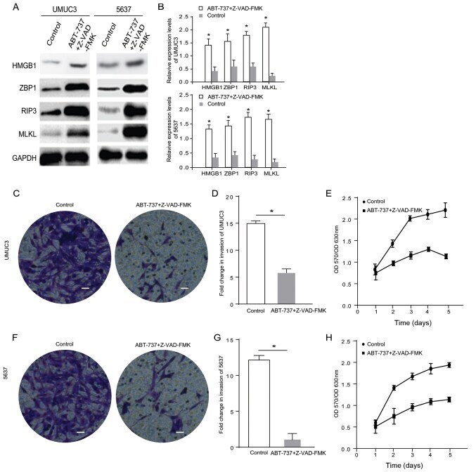
- Figure 2. ABT-737 inhibits the viability and invasion of bladder cancer cells by inducing cell necrosis. (A) Expression levels of HMGB1, ZBP1, RIP3 and MLKL were analyzed via western blotting and the results were (B) semi-quantified. (C) Transwell invasion assays were performed with UMUC3 cells treated with Z-VAD-FMK combined with ABT-737 for 12 h. (D) Quantification of Transwell assay results (E) MTT assays were performed to examine the viability of UMUC3 cells treated with-VAD-FMK combined with ABT-737 for 12 h. (F) Transwell invasion assays were performed with 5637 cells treated with Z-VAD-FMK combined with ABT-737 for 12 h. (G) Quantification of Transwell assay results (H) MTT assays were performed to examine the viability of 5637 cells treated with Z-VAD-FMK combined with ABT-737 for 12 h. (scale bar=0.5 um). *P
- Submitted by
- Invitrogen Antibodies (provider)
- Main image
- Experimental details
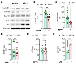
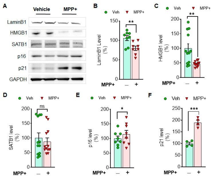
- Figure 1 MPP + toxicity reduces the levels of Lamin B1 and HMGB1, while it increases a cell arrester, p21 in N27 cells. The examples of cellular senescence markers are displayed in Western blots ( A ). The levels of LaminB1 ( B ), HMGB1 ( C ), SATB1 ( D ), p16 ( E ) and p21 ( F ) are displayed in comparison between MPP + and vehicle treatment. Cellular senescence markers such as LaminB1 and HMGB1 were reduced, but p16 and p21 were enhanced by MPP + treatment. GAPDH was adopted as a loading control. ImageJ was used for the analyses of band intensities, and relative levels (100% for vehicle) are displayed in mean +- SEM and applied to unpaired Student's t -test for statistical significance. *: p < 0.05, **: p < 0.01 and ***: p < 0.001. ns: not significant.
- Submitted by
- Invitrogen Antibodies (provider)
- Main image

- Experimental details
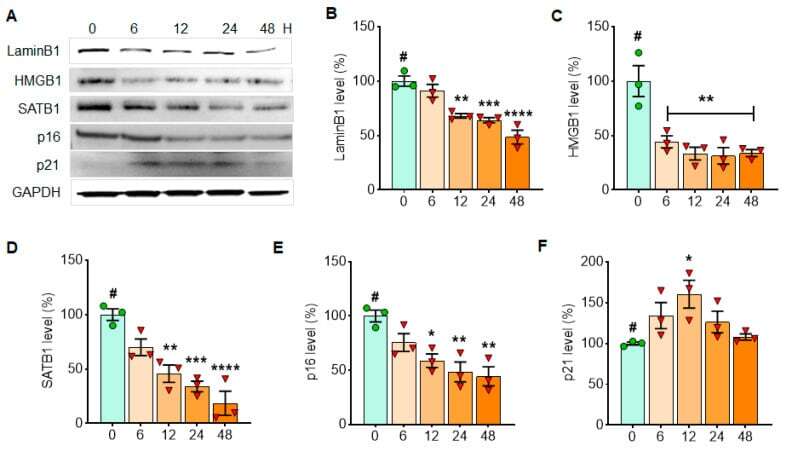
- Figure 2 alpha-Syn PFF treatment reduces the expressions of Lamin B1, HMGB1 and SATB1 in N27 cells, whereas it increases p21 expression in a short period of time. The examples of cellular senescence markers over 48 h are displayed in Western blots ( A ). The levels of LaminB1 ( B ), HMGB1 ( C ), SATB1 ( D ), p16 ( E ) and p21 ( F ) with alpha-syn PFF treatment are displayed in comparison with initial time point (T = 0). GAPDH was adopted as a loading control, and ImageJ was used for the analyses of band intensities. The relative levels (100% for no PFF treated vehicle) are displayed in mean +- SEM and applied to one-way ANOVA, Dunnett's post-hoc test (#: compared with T = 0 point) for statistical significance. * : p < 0.05, ** : p < 0.01, *** : p < 0.001 and **** : p < 0.0001.
- Submitted by
- Invitrogen Antibodies (provider)
- Main image
- Experimental details
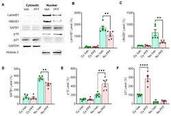
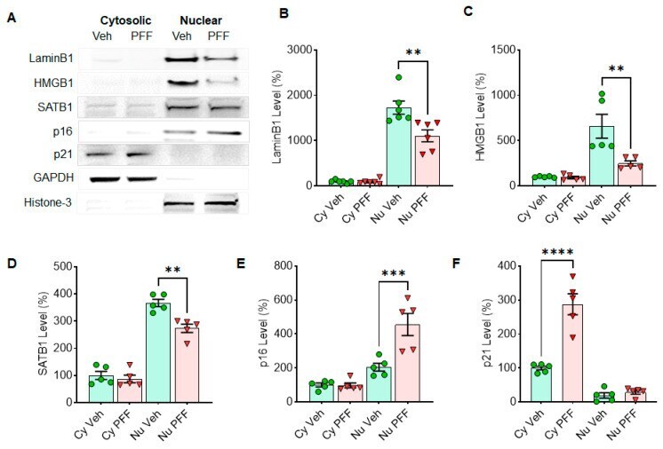
- Figure 3 Senescence markers in N27 cells are regulated in nuclear (Nu) or cytosolic (Cy) fraction by alpha-syn PFF treatment. The alpha-syn PFF treatment reduces the levels of Lamin B1, HMGB1 and SATB1 in nuclear fractions, whereas it increases the levels of p16 in nuclear and p21 in cytosolic fractions after 48 h of exposure. The examples of cellular senescence markers are displayed in Western blots ( A ). The band intensities of Lamin B1 ( B ), HMGB1 ( C ), SATB1 ( D ), p16 ( E ) and p21 ( F ) were quantified using ImageJ. GAPDH and histone-3 were used as a cytosolic- and a nuclear-loading control, respectively. The relative levels (100% for vehicle treatment) are displayed in mean +- SEM and applied to unpaired Student's t -test for statistical significance. ** : p < 0.01, *** : p < 0.001 and **** : p < 0.0001.
- Submitted by
- Invitrogen Antibodies (provider)
- Main image
- Experimental details
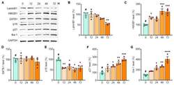
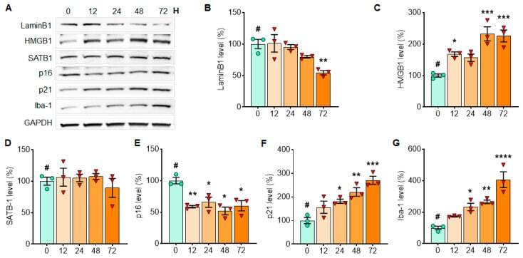
- Figure 6 The levels of LaminB1 and p16 gradually decrease with alpha-syn PFF treatment, while the levels of HMGB1, p21 and Iba-1 increase in the isolated microglial culture over 3 days of alpha-syn PFF exposure. In Western blots, senescence markers were quantified in primary microglia culture at 12, 24, 48 and 72 h after alpha-syn PFF exposure to detect expression patterns over time ( A ). For assessing the expression levels over 3 days of alpha-syn PFF exposure, we quantified the levels of Lamin B1 ( B ), HMGB1 ( C ), SATB1 ( D ), p16 ( E ), p21 ( F ) and Iba-1 ( G ). GAPDH was also used as a loading control. The relative band intensities (100% for no PFF treatment) are displayed in mean +- SEM and applied to one-way ANOVA, Dunnett's post-hoc test (#: compared with T = 0 point) for statistical significance. * : p < 0.05, ** : p < 0.01, *** : p < 0.001 and **** : p < 0.0001.
- Submitted by
- Invitrogen Antibodies (provider)
- Main image
- Experimental details
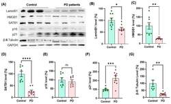
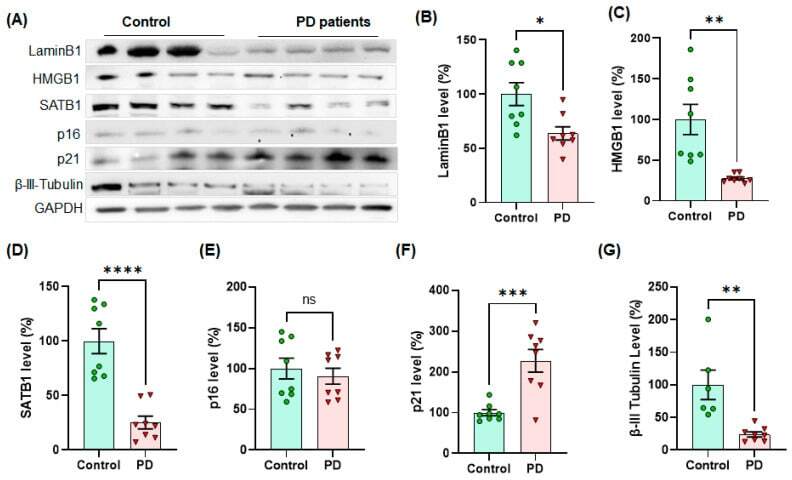
- Figure 7 The levels of cellular senescence markers, such as Lamin B1, HMGB1 and SATB1, were significantly lower in human PD SNpc than age- and gender-matched control tissues, whereas the level of p21 was higher in human PD post-mortem SNpc (n = 8/group). In Western blots, the examples of senescence markers are displayed in quadruplets per group ( A ). The quantified levels of Lamin B1 ( B ), HMGB1 ( C ), SATB1 ( D ), p16 ( E ), p21 ( F ) and beta-III-tubulin ( G ) were statistically analyzed in unpaired Student's t -test for significance. The band intensity of GAPDH was normalized and displayed as relative band intensities (100% for age-matched controls, n = 8/group) in mean +- SEM. * : p < 0.05, ** : p < 0.01, *** : p < 0.001 and **** : p < 0.0001. ns: not significant.
- Submitted by
- Invitrogen Antibodies (provider)
- Main image
- Experimental details
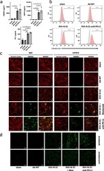
- Induction of immunogenic cell death by XVir-N-31 and XVir-N-31-anti-PD-L1. ( a ) U87MG cells were infected with 50 MOI XVir-N-31 or XVir-N-31-anti-PD-L1, or with 20 MOI Ad-WT, or were left untreated. Supernatants were taken at the timepoint the cultures showed 50% oncolysis, and were analyzed for HMGB1, HSP70, or YB-1 release by ELISA (XVir-PD-L1: XVir-N-31-anti-PD-L1). ( b ) CRT surface expression was analyzed in the U87MG cells at the same conditions as indicated in A (n = 3; SEM; * p < 0.05; ** p < 0.01). ( c ) The detection of HMGB1 in U87MG ipsilateral virus-injected and contralateral tumors 35 days after intratumoral injection of either PBS (sham), 3 x 10 8 IFU of either Ad-WT, of XVir-N-31 alone or in combination with multiple systemic applications of nivolumab, or of XVir-N-31-anti-PD-L1 (n = 7-8 mice per group; representative pictures are shown). ( d ) Enlightenment of the HMGB1 staining as indicated in ( c ).