Explore
 Validate
 Learn
LearnM00066-1
antibody from Boster Biological Technology
Targeting: HMGB1
DKFZp686A04236, HMG1, HMG3, SBP-1
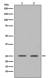
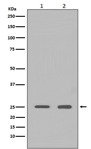
 Western blot
 Immunohistochemistry
Antibody data
- Antibody Data
- Antigen structure
- References [12]
- Comments [0]
- Validations
- Immunohistochemistry [1]
Submit
Validation data
Reference
Comment
Report error
- Product number
- M00066-1 - Provider product page

- Provider
- Boster Biological Technology
- Product name
- Anti-HMGB1/Hmg 1 Rabbit Monoclonal Antibody
- Antibody type
- Monoclonal
- Description
- Monoclonal antibody for HMG 1/HMGB1 detection. Host: Rabbit.Size: 100ug/vial. Tested applications: Flow Cytometry, IF, IHC, ICC, WB. Reactive species: Human, Mouse, Rat HMG 1/HMGB1 information: Molecular Weight: 24894 MW; Subcellular Localization: Nucleus . Chromosome . Cytoplasm . Secreted . Cell membrane ; Peripheral membrane protein ; Extracellular side . Endosome . Endoplasmic reticulum-Golgi intermediate compartment . In basal state predominantly nuclear. Shuttles between the cytoplasm and the nucleus (PubMed:12231511, PubMed:17114460). Translocates from the nucleus to the cytoplasm upon autophagy stimulation (PubMed:20819940). Release from macrophages in the extracellular milieu requires the activation of NLRC4 or NLRP3 inflammasomes (By similarity). Passively released to the extracellular milieu from necrotic cells by diffusion, involving the fully reduced HGMB1 which subsequently gets oxidized (PubMed:19811284). Also released from apoptic cells (PubMed:16855214, PubMed:18631454). Active secretion from a variety of immune and non-immune cells such as macrophages, monocytes, neutrophils, dendritic cells and natural killer cells in response to va
- Reactivity
- Human, Mouse, Rat
- Host
- Rabbit
- Antibody clone number
- AAC-8
- Vial size
- 100ug/vial
- Concentration
- 0.5-1mg/ml, actual concentration vary by lot. Use suggested dilution ratio to decide dilution procedure.
- Storage
- At -20°C for one year. Avoid repeated freezing and thawing.
Submitted references n-3 polyunsaturated fatty acids alleviate the progression of obesity-related osteoarthritis and protect cartilage through inhibiting the HMGB1-RAGE/TLR4 signaling pathway.
Urolithin A protects against acetaminophen-induced liver injury in mice via sustained activation of Nrf2.
Short-term pretreatment of naringin isolated from Citrus wilsonii Tanaka attenuates rat myocardial ischemia/reperfusion injury.
Citrus fruits are rich in flavonoids for immunoregulation and potential targeting ACE2.
HMGB1 in the mPFC governs comorbid anxiety in neuropathic pain.
Abdominal paracentesis drainage attenuates intestinal inflammation in rats with severe acute pancreatitis by inhibiting the HMGB1-mediated TLR4 signaling pathway.
Abdominal paracentesis drainage ameliorates myocardial injury in severe experimental pancreatitis rats through suppressing oxidative stress.
H19 promote calcium oxalate nephrocalcinosis-induced renal tubular epithelial cell injury via a ceRNA pathway.
High-Mobility Group Box 1 Protein Regulates Autophagy in LO2 Cells Following Anoxia-Reoxygenation Injury.
The HMGB1‑IL‑17A axis contributes to hypoxia/reoxygenation injury via regulation of cardiomyocyte apoptosis and autophagy.
Propofol inhibits the release of interleukin-6, 8 and tumor necrosis factor-α correlating with high-mobility group box 1 expression in lipopolysaccharides-stimulated RAW 264.7 cells.
MicroRNA-205‑5b inhibits HMGB1 expression in LPS-induced sepsis.
Xiong T, Huang S, Wang X, Shi Y, He J, Yuan Y, Wang R, Gu H, Liu L
International immunopharmacology 2024 Feb 15;128:111498
International immunopharmacology 2024 Feb 15;128:111498
Urolithin A protects against acetaminophen-induced liver injury in mice via sustained activation of Nrf2.
Gao Z, Yi W, Tang J, Sun Y, Huang J, Lan T, Dai X, Xu S, Jin ZG, Wu X
International journal of biological sciences 2022;18(5):2146-2162
International journal of biological sciences 2022;18(5):2146-2162
Short-term pretreatment of naringin isolated from Citrus wilsonii Tanaka attenuates rat myocardial ischemia/reperfusion injury.
Liu W, Cheng L, Li X, Zhao L, Hu X, Ma Z
Naunyn-Schmiedeberg's archives of pharmacology 2022 Sep;395(9):1047-1059
Naunyn-Schmiedeberg's archives of pharmacology 2022 Sep;395(9):1047-1059
Citrus fruits are rich in flavonoids for immunoregulation and potential targeting ACE2.
Liu W, Zheng W, Cheng L, Li M, Huang J, Bao S, Xu Q, Ma Z
Natural products and bioprospecting 2022 Feb 14;12(1):4
Natural products and bioprospecting 2022 Feb 14;12(1):4
HMGB1 in the mPFC governs comorbid anxiety in neuropathic pain.
Du Y, Xu CL, Yu J, Liu K, Lin SD, Hu TT, Qu FH, Guo F, Lou GD, Nishibori M, Hu WW, Chen Z, Zhang SH
The journal of headache and pain 2022 Aug 16;23(1):102
The journal of headache and pain 2022 Aug 16;23(1):102
Abdominal paracentesis drainage attenuates intestinal inflammation in rats with severe acute pancreatitis by inhibiting the HMGB1-mediated TLR4 signaling pathway.
Huang SQ, Wen Y, Sun HY, Deng J, Zhang YL, Huang QL, Wang B, Luo ZL, Tang LJ
World journal of gastroenterology 2021 Mar 7;27(9):815-834
World journal of gastroenterology 2021 Mar 7;27(9):815-834
Abdominal paracentesis drainage ameliorates myocardial injury in severe experimental pancreatitis rats through suppressing oxidative stress.
Wen Y, Sun HY, Tan Z, Liu RH, Huang SQ, Chen GY, Qi H, Tang LJ
World journal of gastroenterology 2020 Jan 7;26(1):35-54
World journal of gastroenterology 2020 Jan 7;26(1):35-54
H19 promote calcium oxalate nephrocalcinosis-induced renal tubular epithelial cell injury via a ceRNA pathway.
Liu H, Ye T, Yang X, Liu J, Jiang K, Lu H, Xia D, Peng E, Chen Z, Sun F, Tang K, Ye Z
EBioMedicine 2019 Dec;50:366-378
EBioMedicine 2019 Dec;50:366-378
High-Mobility Group Box 1 Protein Regulates Autophagy in LO2 Cells Following Anoxia-Reoxygenation Injury.
Li M, Peng G, Ye Q, Wang Y, Xiong Y, Wang R, Yang Z
Transplantation proceedings 2018 Jun;50(5):1532-1537
Transplantation proceedings 2018 Jun;50(5):1532-1537
The HMGB1‑IL‑17A axis contributes to hypoxia/reoxygenation injury via regulation of cardiomyocyte apoptosis and autophagy.
Hu X, Zhang K, Chen Z, Jiang H, Xu W
Molecular medicine reports 2018 Jan;17(1):336-341
Molecular medicine reports 2018 Jan;17(1):336-341
Propofol inhibits the release of interleukin-6, 8 and tumor necrosis factor-α correlating with high-mobility group box 1 expression in lipopolysaccharides-stimulated RAW 264.7 cells.
Jia J, Sun Y, Hu Z, Li Y, Ruan X
BMC anesthesiology 2017 Oct 26;17(1):148
BMC anesthesiology 2017 Oct 26;17(1):148
MicroRNA-205‑5b inhibits HMGB1 expression in LPS-induced sepsis.
Zhou W, Wang J, Li Z, Li J, Sang M
International journal of molecular medicine 2016 Jul;38(1):312-8
International journal of molecular medicine 2016 Jul;38(1):312-8
No comments: Submit comment
Supportive validation
- Submitted by
- Boster Biological Technology (provider)
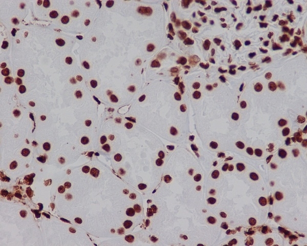
- Main image
- Experimental details
- Immunohistochemical analysis of paraffin-embedded human kidney, using HMGB1 Antibody(M00066-1)HMGB1 was detected in paraffin-embedded tissue section. Heat mediated antigen retrieval was performed in citrate buffer (pH6, epitope retrieval solution) for 20 mins. The tissue section was blocked with 10% goat serum. The tissue section was then incubated with 1ug/ml rabbit anti-HMGB1 Antibody (M00066-1)overnight at 4°C. Biotinylated goat anti-rabbit IgG was used as secondary antibody and incubated for 30 minutes at 37°C. The tissue section was developed using Strepavidin-Biotin-Complex (SABC)(Catalog # SA1022) with DAB as the chromogen.
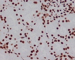
- Additional image