Explore
 Validate
 Learn
Learn Western blot
Antibody data
- Antibody Data
- Antigen structure
- References [0]
- Comments [0]
- Validations
- Western blot [1]
- Immunohistochemistry [4]
- Flow cytometry [2]
Submit
Validation data
Reference
Comment
Report error
- Product number
- 10-4024 - Provider product page

- Provider
- ABEOMICS Inc.
- Product name
- Anti-HMGB1 Antibody
- Antibody type
- Monoclonal
- Description
- HMGB1 (High-mobility group protein B1) is a 25 kDa non-histone chromatin protein, secreted by immune cells through leaderless secretory pathway. HMGB1 supports transcription and also interacts with nucleosomes to loosen packed DNA and remodel the chromatin. The presence of HMGB1 in the nucleus depends on posttranslational modifications. When the protein is not acetylated, it stays in the nucleus, but hyperacetylation on lysine residues causes it to translocate into the cytosol. HMGB1 has the ability to bend DNA in order to facilitate binding of other proteins to the DNA. HMGB1 has one important role in V(D)J recombination where act as a cofactor of the RAG complex. HMGB1 stimulates a plethora of cellular responses like inflammation, attracting inflammatory cells, recruiting stem cells and promoting their proliferation. HMGB1 is highly expressed in human kidney, tonsils, adipose tissue, adrenal cortex and amniotic fluid.
- Reactivity
- Human
- Host
- Mouse
- Conjugate
- Unconjugated
- Antigen sequence
A partial length recombinant HMGB1
protein (amino acids 1- 200) was us
ed as the immunogen for this antibo
dy.- Isotype
- IgG
- Antibody clone number
- ABM24D3
- Vial size
- 100 µg
- Concentration
- 0.5 mg/ml
- Storage
- Store the antibody at 4°C, stable for 6 months. For long-term storage, store at -20°C. Avoid repeat freez thawing
No comments: Submit comment
Supportive validation
- Submitted by
- ABEOMICS Inc. (provider)
- Main image
- Experimental details
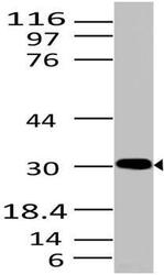
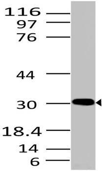
- Western blot analysis of HMGB1. Anti- HMGB1 antibody (Clone: ABM24D3) was used at 2 µg/ml on HepG2 lysate.
- Protocol
- Protocol
Supportive validation
- Submitted by
- ABEOMICS Inc. (provider)
- Main image

- Experimental details
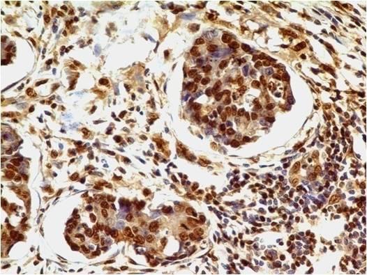
- Immunohistochemical analysis of HMGB1 in adenocarcinoma of stomach using HMGB1 antibody (Clone: ABM24D3) at 1 µg/ml.
- Protocol
- Protocol
- Submitted by
- ABEOMICS Inc. (provider)
- Main image
- Experimental details
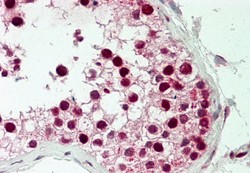
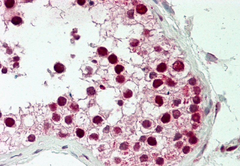
- Immunohistochemical analysis of HMGB1 in human Testis tissue using HMGB1 antibody (Clone: ABM24D3) at 5 µg/ml.
- Protocol
- Protocol
- Submitted by
- ABEOMICS Inc. (provider)
- Main image

- Experimental details
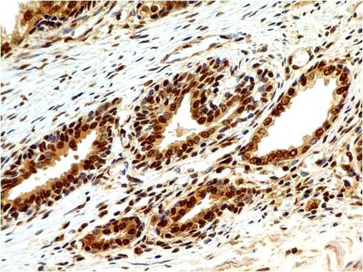
- Immunohistochemical analysis of HMGB1 in human Prostate using HMGB1 antibody (Clone: ABM24D3) at 1 µg/ml.
- Protocol
- Protocol
- Submitted by
- ABEOMICS Inc. (provider)
- Main image
- Experimental details
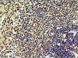
- Immunohistochemical analysis of HMGB1 in human Spleen using HMGB1 antibody (Clone: ABM24D3) at 1 µg/ml.
- Protocol
- Protocol

Supportive validation
- Submitted by
- ABEOMICS Inc. (provider)
- Main image
- Experimental details
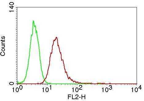
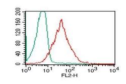
- Intracellular flow analysis of HMGB1 in PBMC (Lymphocyte) using 0.5 µg/10^6 cells of HMGB1 antibody (Clone: ABM24D3). Green represents isotype control; red represents anti-HMGB1 antibody. Goat anti-mouse PE conjugate was used as secondary.
- Protocol
- Protocol
- Submitted by
- ABEOMICS Inc. (provider)
- Main image
- Experimental details
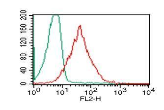
- Intracellular flow analysis of HMGB1 in Jurkat cells using 0.5 µg/10^6 cells of HMGB1 antibody (Clone: ABM24D3). Green represents isotype control; red represents anti-HMGB1 antibody. Goat anti-mouse PE conjugate was used as secondary antibody.
- Protocol
- Protocol