Explore
 Validate
 Learn
Learn Western blot
 ELISA
ELISA Immunocytochemistry
Antibody data
- Antibody Data
- Antigen structure
- References [8]
- Comments [0]
- Validations
- Immunocytochemistry [2]
- Immunohistochemistry [1]
- Flow cytometry [5]
- Chromatin Immunoprecipitation [2]
- Other assay [6]
Submit
Validation data
Reference
Comment
Report error
- Product number
- PA1-16926 - Provider product page

- Provider
- Invitrogen Antibodies
- Product name
- HMGB1 Polyclonal Antibody
- Antibody type
- Polyclonal
- Antigen
- Synthetic peptide
- Description
- This antibody is expected to cross react with porcine, equine and rabbit samples based on 100% sequence homology. Suggested positive control: Hela whole cell extract, antigen standard for HMGB1 (transient overexpression lysate).
- Reactivity
- Human, Mouse, Rat, Bovine, Canine, Rabbit
- Host
- Rabbit
- Isotype
- IgG
- Vial size
- 100 μL
- Concentration
- 1 mg/mL
- Storage
- 4°C, do not freeze
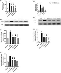
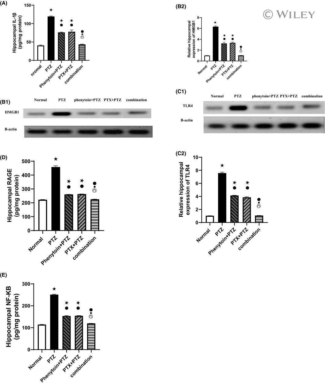
Submitted references Pentoxifylline prevents epileptic seizure via modulating HMGB1/RAGE/TLR4 signalling pathway and improves memory in pentylenetetrazol kindling rats.
The Potential of Nanobody-Targeted Photodynamic Therapy to Trigger Immune Responses.
A small bioactive glycoside inhibits epsilon toxin and prevents cell death.
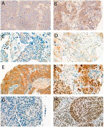
Expression Analysis of Autophagy Related Markers LC3B, p62 and HMGB1 Indicate an Autophagy-Independent Negative Prognostic Impact of High p62 Expression in Pulmonary Squamous Cell Carcinomas.
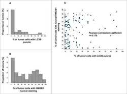
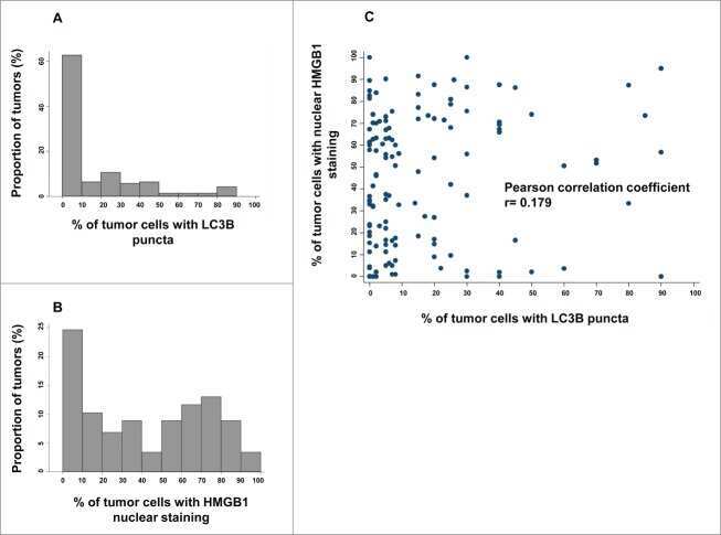
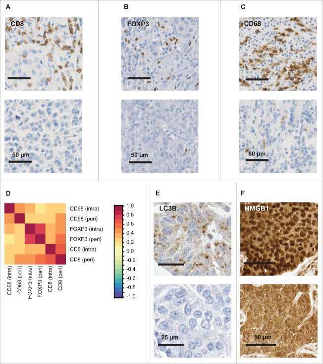
The presence of LC3B puncta and HMGB1 expression in malignant cells correlate with the immune infiltrate in breast cancer.
Biomarkers of immunogenic stress in metastases from melanoma patients: Correlations with the immune infiltrate.
Combined evaluation of LC3B puncta and HMGB1 expression predicts residual risk of relapse after adjuvant chemotherapy in breast cancer.
Negative prognostic impact of regulatory T cell infiltration in surgically resected esophageal cancer post-radiochemotherapy.
Badawi GA, Shokr MM, Zaki HF, Mohamed AF
Clinical and experimental pharmacology & physiology 2021 Aug;48(8):1111-1124
Clinical and experimental pharmacology & physiology 2021 Aug;48(8):1111-1124
The Potential of Nanobody-Targeted Photodynamic Therapy to Trigger Immune Responses.
Beltrán Hernández I, Angelier ML, Del Buono D'Ondes T, Di Maggio A, Yu Y, Oliveira S
Cancers 2020 Apr 15;12(4)
Cancers 2020 Apr 15;12(4)
A small bioactive glycoside inhibits epsilon toxin and prevents cell death.
Shivappagowdar A, Pati S, Narayana C, Ayana R, Kaushik H, Sah R, Garg S, Khanna A, Kumari J, Garg L, Sagar R, Singh S
Disease models & mechanisms 2019 Oct 10;12(10)
Disease models & mechanisms 2019 Oct 10;12(10)
Expression Analysis of Autophagy Related Markers LC3B, p62 and HMGB1 Indicate an Autophagy-Independent Negative Prognostic Impact of High p62 Expression in Pulmonary Squamous Cell Carcinomas.
Langer R, Neppl C, Keller MD, Schmid RA, Tschan MP, Berezowska S
Cancers 2018 Aug 21;10(9)
Cancers 2018 Aug 21;10(9)
The presence of LC3B puncta and HMGB1 expression in malignant cells correlate with the immune infiltrate in breast cancer.
Ladoire S, Enot D, Senovilla L, Ghiringhelli F, Poirier-Colame V, Chaba K, Semeraro M, Chaix M, Penault-Llorca F, Arnould L, Poillot ML, Arveux P, Delaloge S, Andre F, Zitvogel L, Kroemer G
Autophagy 2016 May 3;12(5):864-75
Autophagy 2016 May 3;12(5):864-75
Biomarkers of immunogenic stress in metastases from melanoma patients: Correlations with the immune infiltrate.
Ladoire S, Senovilla L, Enot D, Ghiringhelli F, Poirier-Colame V, Chaba K, Erdag G, Schaefer JT, Deacon DH, Zitvogel L, Slingluff CL Jr, Kroemer G
Oncoimmunology 2016 Jun;5(6):e1160193
Oncoimmunology 2016 Jun;5(6):e1160193
Combined evaluation of LC3B puncta and HMGB1 expression predicts residual risk of relapse after adjuvant chemotherapy in breast cancer.
Ladoire S, Penault-Llorca F, Senovilla L, Dalban C, Enot D, Locher C, Prada N, Poirier-Colame V, Chaba K, Arnould L, Ghiringhelli F, Fumoleau P, Spielmann M, Delaloge S, Poillot ML, Arveux P, Goubar A, Andre F, Zitvogel L, Kroemer G
Autophagy 2015;11(10):1878-90
Autophagy 2015;11(10):1878-90
Negative prognostic impact of regulatory T cell infiltration in surgically resected esophageal cancer post-radiochemotherapy.
Vacchelli E, Semeraro M, Enot DP, Chaba K, Poirier Colame V, Dartigues P, Perier A, Villa I, Rusakiewicz S, Gronnier C, Goéré D, Mariette C, Zitvogel L, Kroemer G
Oncotarget 2015 Aug 28;6(25):20840-50
Oncotarget 2015 Aug 28;6(25):20840-50
No comments: Submit comment
Supportive validation
- Submitted by
- Invitrogen Antibodies (provider)
- Main image

- Experimental details
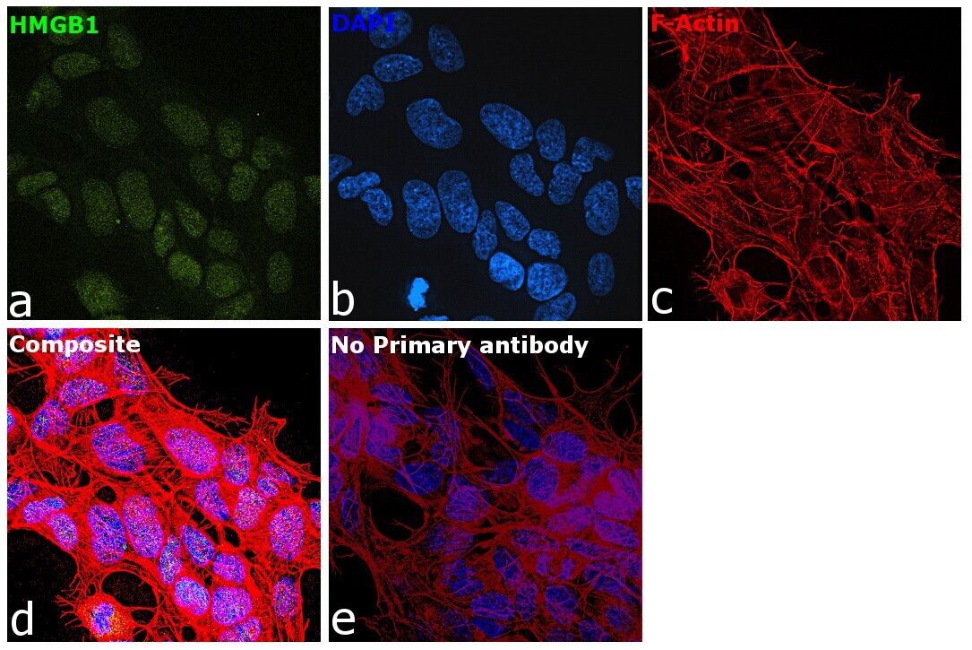
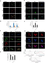
- Immunofluorescence analysis of HMGB1 was performed using 70% confluent log phase SH-SY5Y cells. The cells were fixed with 4% paraformaldehyde for 10 minutes, permeabilized with 0.1% Triton™ X-100 for 15 minutes, and blocked with 2% BSA for 45 minutes at room temperature. The cells were labeled with HMGB1 Polyclonal Antibody (Product # PA1-16926) at 0.05 µg/mL in 0.1% BSA, incubated at 4 degree celsius overnight and then labeled with Donkey anti-Rabbit IgG (H+L) Highly Cross-Adsorbed Secondary Antibody, Alexa Fluor Plus 488 (Product # A32790), (1:2000), for 45 minutes at room temperature (Panel a: Blue). Nuclei (Panel b:Green) were stained with ProLong™ Diamond Antifade Mountant with DAPI (Product # P36962). F-actin (Panel c: Red) was stained with Rhodamine Phalloidin (Product # R415, 1:300). Panel d represents the merged image showing Nuclear localization. Panel e represents control cells with no primary antibody to assess background. The images were captured at 60X magnification.

- Submitted by
- Invitrogen Antibodies (provider)
- Main image

- Experimental details
- Immunofluorescence analysis of HMGB1 was performed using 70% confluent log phase SH-SY5Y cells. The cells were fixed with 4% paraformaldehyde for 10 minutes, permeabilized with 0.1% Triton™ X-100 for 15 minutes, and blocked with 2% BSA for 45 minutes at room temperature. The cells were labeled with HMGB1 Polyclonal Antibody (Product # PA1-16926) at 0.05 µg/mL in 0.1% BSA, incubated at 4 degree celsius overnight and then labeled with Donkey anti-Rabbit IgG (H+L) Highly Cross-Adsorbed Secondary Antibody, Alexa Fluor Plus 488 (Product # A32790), (1:2000), for 45 minutes at room temperature (Panel a: Blue). Nuclei (Panel b:Green) were stained with ProLong™ Diamond Antifade Mountant with DAPI (Product # P36962). F-actin (Panel c: Red) was stained with Rhodamine Phalloidin (Product # R415, 1:300). Panel d represents the merged image showing Nuclear localization. Panel e represents control cells with no primary antibody to assess background. The images were captured at 60X magnification.
Supportive validation
- Submitted by
- Invitrogen Antibodies (provider)
- Main image
- Experimental details
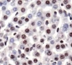
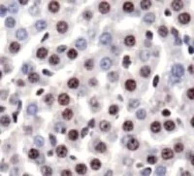
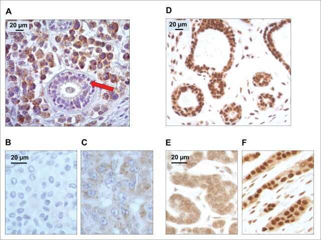
- Immunohistochemical analysis of HMGB1 in mouse liver. Samples were incubated in HMGB1 polyclonal antibody (Product # PA1-16926).
Supportive validation
- Submitted by
- Invitrogen Antibodies (provider)
- Main image
- Experimental details
- FACS staining of NTERA-2 cells using Product # PA1-16926 at a 1:50 dilution detected using Dylight-488 conjugated goat anti-rabbit IgG secondary antibody.
- Submitted by
- Invitrogen Antibodies (provider)
- Main image
- Experimental details
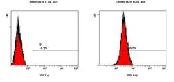
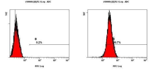
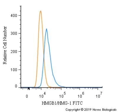
- Flow cytometry of HMGB1 in HeLa and a matched isotype control. Samples were incubated in HMGB1 polyclonal antibody (Product # PA1-16926) using a dilution of 1 µg/mL for 30 minutes at room temperature followed by a Rabbit IgG (H+L) Cross-Adsorbed secondary antibody. Cells were fixed with 4% PFA and then permeabilized with 0.1% saponin.

- Submitted by
- Invitrogen Antibodies (provider)
- Main image

- Experimental details
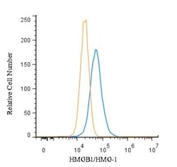
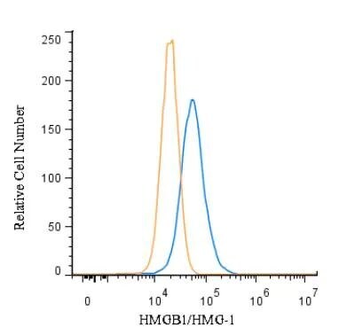
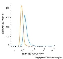
- Flow cytometry of HMGB1 in RH-30 cells. Samples were incubated in HMGB1 polyclonal antibody (Product # PA1-16926) using a dilution of 10 µg/mL for 30 minutes at room temperature. Antibody (blue) and a matched isotype control (orange). Cells were fixed with 4% PFA and then permeabilized with 0.1% saponin.

- Submitted by
- Invitrogen Antibodies (provider)
- Main image

- Experimental details
- Flow cytometry of HMGB1 in HeLa and a matched isotype control. Samples were incubated in HMGB1 polyclonal antibody (Product # PA1-16926) using a dilution of 1 µg/mL for 30 minutes at room temperature followed by a Rabbit IgG (H+L) Cross-Adsorbed secondary antibody. Cells were fixed with 4% PFA and then permeabilized with 0.1% saponin.
- Submitted by
- Invitrogen Antibodies (provider)
- Main image
- Experimental details
- Flow cytometry of HMGB1 in RH-30 cells. Samples were incubated in HMGB1 polyclonal antibody (Product # PA1-16926) using a dilution of 10 µg/mL for 30 minutes at room temperature. Antibody (blue) and a matched isotype control (orange). Cells were fixed with 4% PFA and then permeabilized with 0.1% saponin.
Supportive validation
- Submitted by
- Invitrogen Antibodies (provider)
- Main image

- Experimental details
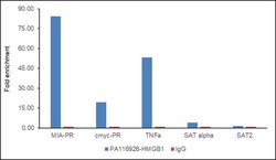
- Chromatin Immunoprecipitation (ChIP) assay of endogenous HMGB1 protein using HMGB1 Antibody: ChIP was performed using HMGB1 Polyclonal Antibody (Product # PA1-16926, 5 µg) on sheared chromatin from Hep G2 cells using the MAGnify ChIP System kit (Product # 49-2024). Normal Rabbit IgG was used as a negative IP control. The purified DNA was analyzed by qPCR using primers binding to MIA-PR (promoter), c myc promoter, TNF alpha (active) and SAT alpha and SAT2 satellite repeats (Inactive). Data is presented as fold enrichment of the antibody signal versus the negative control IgG using the comparative CT method.

- Submitted by
- Invitrogen Antibodies (provider)
- Main image
- Experimental details
- Chromatin Immunoprecipitation (ChIP) assay of endogenous HMGB1 protein using HMGB1 Antibody: ChIP was performed using HMGB1 Polyclonal Antibody (Product # PA1-16926, 5 µg) on sheared chromatin from Hep G2 cells using the MAGnify ChIP System kit (Product # 49-2024). Normal Rabbit IgG was used as a negative IP control. The purified DNA was analyzed by qPCR using primers binding to MIA-PR (promoter), c myc promoter, TNF alpha (active) and SAT alpha and SAT2 satellite repeats (Inactive). Data is presented as fold enrichment of the antibody signal versus the negative control IgG using the comparative CT method.

Supportive validation
- Submitted by
- Invitrogen Antibodies (provider)
- Main image
- Experimental details
- NULL
- Submitted by
- Invitrogen Antibodies (provider)
- Main image
- Experimental details
- NULL
- Submitted by
- Invitrogen Antibodies (provider)
- Main image
- Experimental details
- NULL
- Submitted by
- Invitrogen Antibodies (provider)
- Main image
- Experimental details
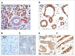
- Figure 1 Examples of immunohistochemical stainings; ( A ) LC3B low, ( B ) LC3B high, ( C ) p62 low (any cellular compartment), ( D ) p62 cytoplasmic low, ( E ) p62 cytoplasmic/dot like high, nuclear low, ( F ) p62 cytoplasmic/dot like low, nuclear high, ( G ) HMGB1 low, ( H ) HMGB1 high. Objective magnification, 40x.

- Submitted by
- Invitrogen Antibodies (provider)
- Main image
- Experimental details
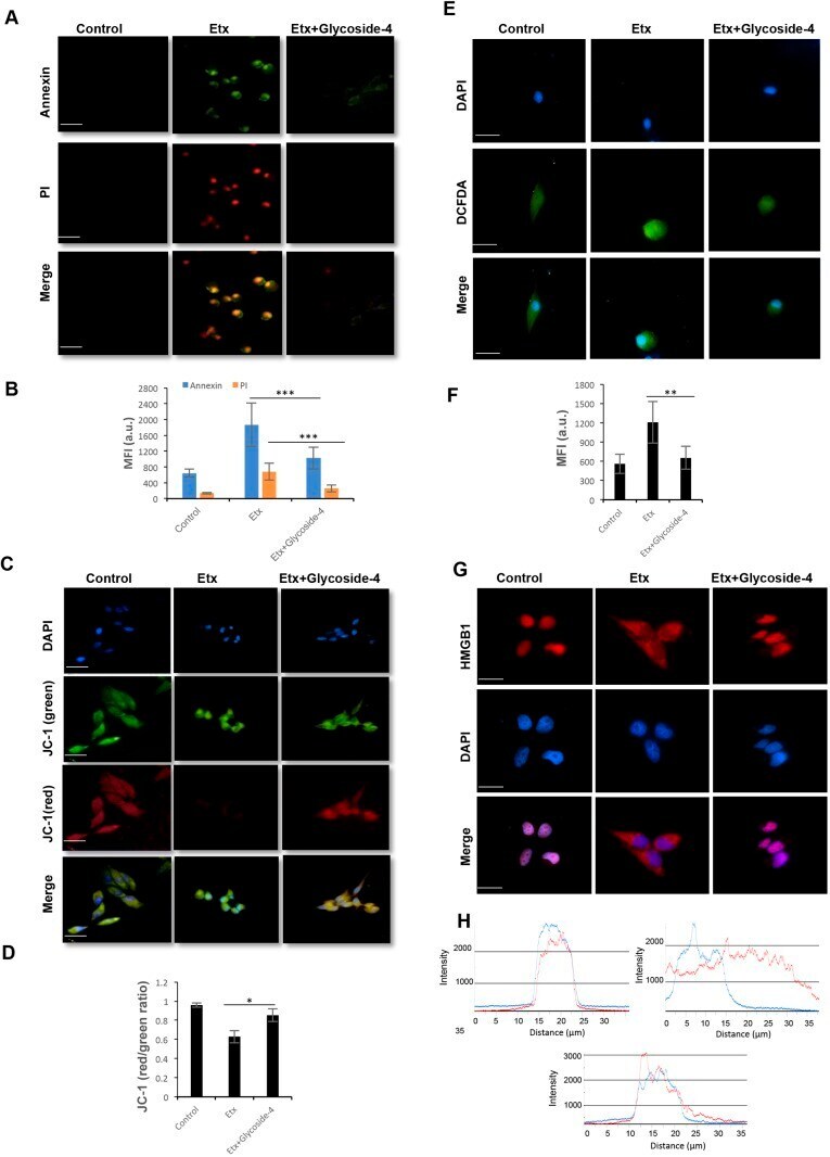
- 4 FIGURE Effect of pentoxifylline (PTX) on pro-inflammatory parameters (IL-1beta, HMGb1, TLR4, RAGE, and NF-kappaB) in hippocampi of pentylenetetrazol (PTZ)-kindled rats. (A): IL-1beta, (B); HMGB1, (C); TLR4, (D); RAGE, (E); NF-kappaB. (B1); Representative Western blot analysis of hippocampal HMGB1, (B2); Quantification of the level of hippocampal HMGB1. (C1); Representative Western blot analysis of hippocampal TLR4, (C2); Quantification of the level of hippocampal TLR4. Data are expressed as mean +- SEM of six rats of each group; p
- Submitted by
- Invitrogen Antibodies (provider)
- Main image
- Experimental details
- Fig. 4. Glycoside-4 protects MDCK cells from Etx-triggered cell death. (A) MDCK cells were treated with Etx or in combination with glycoside-4, and the annexin V staining of exposed phosphatidyl serine on the outer surface and PI entry were observed. The toxin-treated cells showed annexin and PI (annexin + /PI + ) dual positivity, whereas glycoside-4-treated cells showed the absence of both the markers (annexin - /PI - ). Scale bars: 20 mum. (B) Bar graph depicts mean fluorescence intensity (MFI) for annexin and PI. Statistical significance (*** P