Explore
 Validate
 Learn
Learn Western blot
Antibody data
- Antibody Data
- Antigen structure
- References [0]
- Comments [0]
- Validations
- Western blot [2]
- Immunocytochemistry [1]
Submit
Validation data
Reference
Comment
Report error
- Product number
- Ab156561 - Provider product page

- Provider
- Aladdin Scientific
- Product name
- HMGB1 Mouse mAb
- Antibody type
- Monoclonal
- Description
- ApplicationWB: 1/2,000 - 1/100,000; Predicted molecular weight: 25.0 kDaFlow: 0.02 _g/mL - 2.5 _g/mL
- Reactivity
- Human, Mouse, Rat
- Host
- Mouse
- Conjugate
- Unconjugated
- Isotype
- IgG
- Antibody clone number
- AB02/4A1
- Vial size
- 100_l,10_l,1ml,20_l,50_l
- Concentration
- Lot by lot
- Storage
- Store at +4¡C short term (1-2 weeks). Store at -20¡C or -80¡C. Upon delivery aliquot. Avoid freeze/thaw cycle.
No comments: Submit comment
Supportive validation
- Submitted by
- Aladdin Scientific (provider)
- Main image
- Experimental details
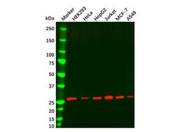
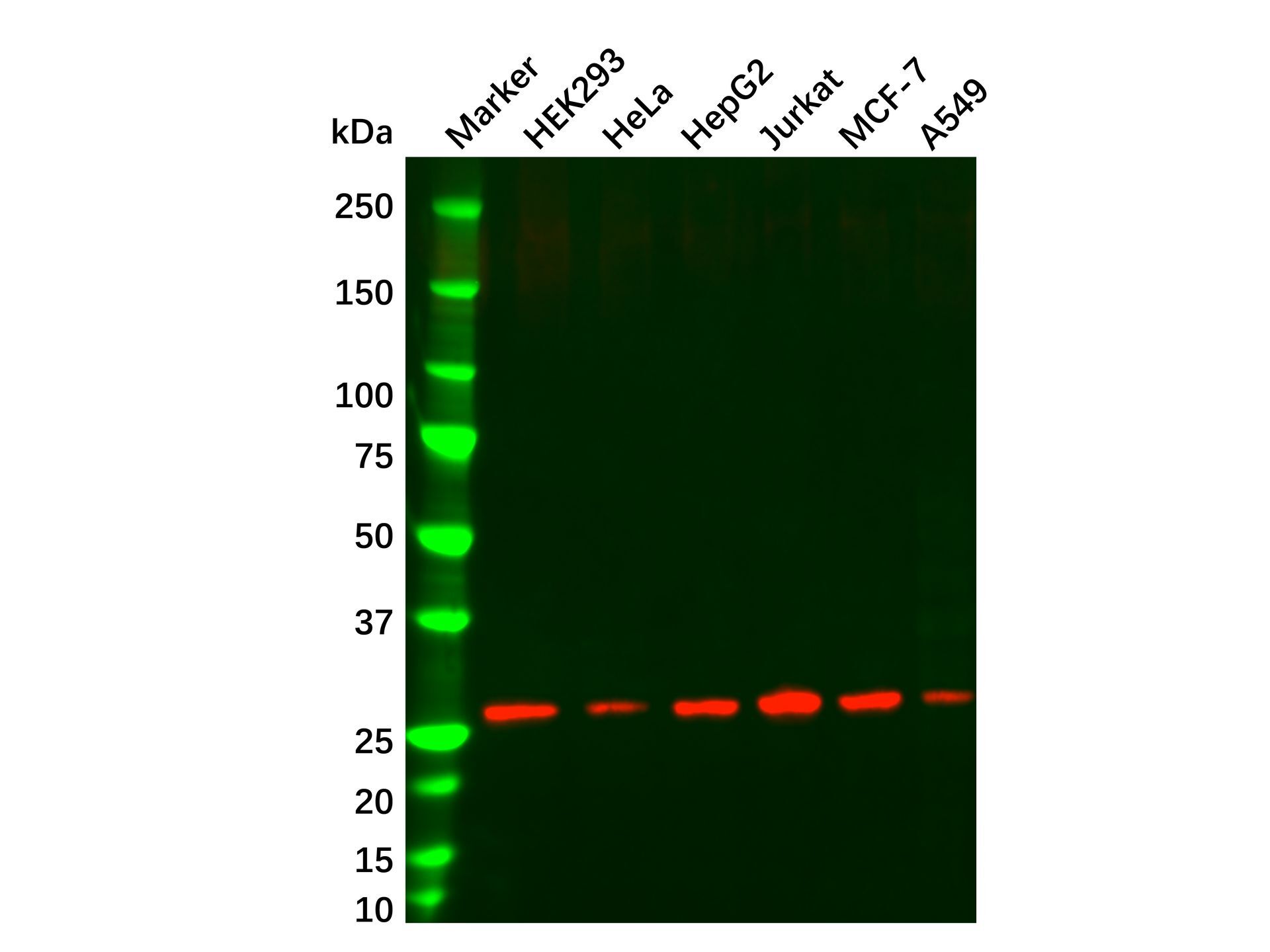
- HMGB1 Mouse mAb (Ab156561) - Western Blot All lanes: HMGB1 Antibody (Ab156561) at 0.1 μg/mL Samples: Lysates at 20 µg per lane Secondary: Goat Anti-Mouse IgG H&L (HRP) at 1/20000 dilution Predicted band size: 24.9 kDa Observed band size: 27.2 kDa Exposure time: 118.1 s
- Submitted by
- Aladdin Scientific (provider)
- Main image
- Experimental details
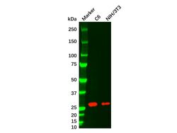
- HMGB1 Mouse mAb (Ab156561) - Western Blot All lanes: HMGB1 Antibody (Ab156561) at 0.1 μg/mL Samples: Lysates at 20 µg per lane Secondary: Goat Anti-Mouse IgG H&L (HRP) at 1/20000 dilution Predicted band size: 24.9 kDa Observed band size: 26.1 kDa Exposure time: 27.8 s

Supportive validation
- Submitted by
- Aladdin Scientific (provider)
- Main image
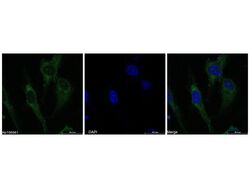
- Experimental details
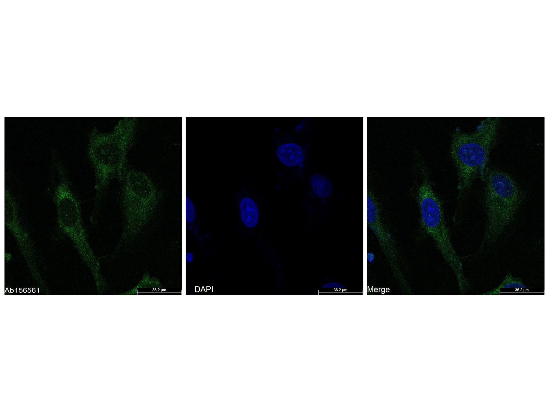
- HMGB1 Mouse mAb (Ab156561) - ICC/IF IF analysis of HMGB1 (Green) in HeLa cells. The cells were fixed and permeabilized with 100% methanol for 5 minutes, and blocked with 2% BSA for 1 hour at room temperature. Cells were stained with HMGB1 Mouse mAb (Ab156561) at 2.0 μg/mL in blocking buffer for 2 hour at RT and then incubated with Goat Anti-Mouse IgG H&L (FITC) (Ab156254) at a dilution of 1/1000 for 1 hour at room temperature in the dark. Cells were counterstained with DAPI (Blue). Images were taken on the confocal laser scanning microscope.