Explore
 Validate
 Learn
Learn Western blot
 Immunocytochemistry
Antibody data
- Antibody Data
- Antigen structure
- References [0]
- Comments [0]
- Validations
- Western blot [5]
- Immunocytochemistry [2]
- Immunoprecipitation [1]
- Immunohistochemistry [4]
Submit
Validation data
Reference
Comment
Report error
- Product number
- GTX127344 - Provider product page

- Provider
- GeneTex
- Proper citation
- GeneTex Cat#GTX127344, RRID:AB_11164700
- Product name
- HMGB1 antibody
- Antibody type
- Polyclonal
- Reactivity
- Human, Mouse, Rat
- Host
- Rabbit
No comments: Submit comment
Enhanced validation
Supportive validation
- Submitted by
- GeneTex (provider)
- Enhanced method
- Genetic validation
- Main image

- Experimental details
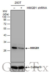
- Non-transfected (¡V) and transfected (+) 293T whole cell extracts (30 ?g) were separated by 12% SDS-PAGE, and the membrane was blotted with HMGB1 antibody (GTX127344) diluted at 1:5000.

Supportive validation
- Submitted by
- GeneTex (provider)
- Main image

- Experimental details
- HMGB1 antibody detects HMGB1 protein by Western blot analysis.A. 30 ?g 293T whole cell lysate/extractB. 30 ?g A431 whole cell lysate/extractC. 30 ?g HeLa whole cell lysate/extractD. 30 ?g HepG2 whole cell lysate/extract12 % SDS-PAGEHMGB1 antibody (GTX127344) dilution: 1:1000

- Submitted by
- GeneTex (provider)
- Main image

- Experimental details
- Mouse tissue extract (50 ?g) was separated by 12% SDS-PAGE, and the membrane was blotted with HMGB1 antibody (GTX127344) diluted at 1:500.

- Submitted by
- GeneTex (provider)
- Main image

- Experimental details
- Rat tissue extract (50 ?g) was separated by 12% SDS-PAGE, and the membrane was blotted with HMGB1 antibody (GTX127344) diluted at 1:500.

- Submitted by
- GeneTex (provider)
- Main image
- Experimental details
- Non-transfected (¡V) and transfected (+) 293T whole cell extracts (30 ?g) were separated by 12% SDS-PAGE, and the membrane was blotted with HMGB1 antibody (GTX127344) diluted at 1:5000.

Supportive validation
- Submitted by
- GeneTex (provider)
- Main image

- Experimental details
- HMGB1 antibody detects HMGB1 protein at nucleus by immunofluorescent analysis.Sample: NT2D1 cells were fixed in 4% paraformaldehyde at RT for 15 min.Green: HMGB1 protein stained by HMGB1 antibody (GTX127344) diluted at 1:500.Blue: Hoechst 33342 staining.Scale bar = 10 £gm.

- Submitted by
- GeneTex (provider)
- Main image

- Experimental details
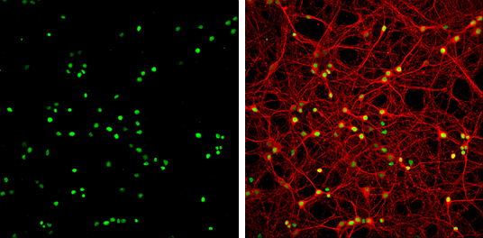
- HMGB1 antibody detects HMGB1 protein at nucleus by immunofluorescent analysis.Sample: DIV9 rat E18 primary cortical neuron cells were fixed in 4% paraformaldehyde at RT for 15 min.Green: HMGB1 protein stained by HMGB1 antibody (GTX127344) diluted at 1:500.Red: beta Tubulin 3/ Tuj1, a neuron cell marker, stained by beta Tubulin 3/ Tuj1 antibody [GT11710] (GTX631836) diluted at 1:500.
Supportive validation
- Submitted by
- GeneTex (provider)
- Main image

- Experimental details
- Immunoprecipitation of HMGB1 protein from 293T whole cell extracts using 5 £gg of HMGB1 antibody (GTX127344).Western blot analysis was performed using HMGB1 antibody (GTX127344).EasyBlot anti-Rabbit IgG (GTX221666-01) was used as a secondary reagent.

Supportive validation
- Submitted by
- GeneTex (provider)
- Main image

- Experimental details
- Immunohistochemical analysis of paraffin-embedded SkHep1 xenograft, using HMGB1(GTX127344) antibody at 1:500 dilution.

- Submitted by
- GeneTex (provider)
- Main image

- Experimental details
- HMGB1 antibody detects HMGB1 protein at nucleus on mouse urinary bladder by immunohistochemical analysis. Sample: Paraffin-embedded mouse urinary bladder. HMGB1 antibody (GTX127344) dilution: 1:500.

- Submitted by
- GeneTex (provider)
- Main image
- Experimental details
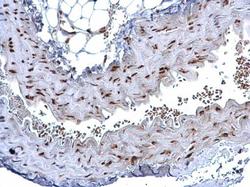
- HMGB1 antibody detects HMGB1 protein at nucleus on mouse vein by immunohistochemical analysis. Sample: Paraffin-embedded mouse vein. HMGB1 antibody (GTX127344) dilution: 1:500.

- Submitted by
- GeneTex (provider)
- Main image

- Experimental details
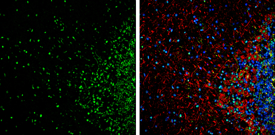
- HMGB1 antibody detects HMGB1 Protein expression by immunohistochemical analysis.Sample: Frozen-sectioned adult mouse cerebellum. Green: HMGB1 stained by HMGB1 antibody (GTX127344) diluted at 1:250.Red: NF-H, stained by NF-H antibody [GT114] (GTX634289) diluted at 1:500.Blue: Fluoroshield with DAPI (GTX30920).