 Explore
 Validate
 Learn
Learn Western blot
Antibody data
- Antibody Data
- Antigen structure
- References [1]
- Comments [0]
- Validations
- Western blot [1]
- Immunohistochemistry [7]
- Flow cytometry [1]
Submit
Validation data
Reference
Comment
Report error
- Product number
- MA5-16264 - Provider product page

- Provider
- Invitrogen Antibodies
- Product name
- HMGB1 Monoclonal Antibody (19N15F4)
- Antibody type
- Monoclonal
- Antigen
- Other
- Reactivity
- Human, Mouse
- Host
- Mouse
- Isotype
- IgG
- Antibody clone number
- 19N15F4
- Vial size
- 100 µg
- Concentration
- 0.5 mg/mL
- Storage
- Store at 4°C short term. For long term storage, store at -20°C, avoiding freeze/thaw cycles.
Submitted references Chronic hyperosmotic stress interferes with immune homeostasis in striped catfish (Pangasianodon hypophthalmus, S.) and leads to excessive inflammatory response during bacterial infection.
Schmitz M, Douxfils J, Mandiki SN, Morana C, Baekelandt S, Kestemont P
Fish & shellfish immunology 2016 Aug;55:550-8
Fish & shellfish immunology 2016 Aug;55:550-8
No comments: Submit comment
Supportive validation
- Submitted by
- Invitrogen Antibodies (provider)
- Main image

- Experimental details
- Western blot analysis of HMGB1 in full-length human HMGB1 protein, human Jurkat cell lysate and mouse NIH 3T3 cell lysate. Samples were incubated in HMGB1 monoclonal antibody (Product # MA5-16264) using a dilution of 2 µg/mL followed by a goat anti-mouse IgG HRP secondary antibody. PicoTect ECL substrate solution was used in this assay.

Supportive validation
- Submitted by
- Invitrogen Antibodies (provider)
- Main image
- Experimental details
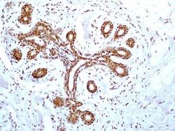
- Immunohistochemical analysis of HMGB1 in a section of human breast normal tissue. Samples were incubated in HMGB1 monoclonal antibody (Product # MA5-16264) using a dilution of 5 µg/mL. The ductal /acinar epithelial cells in the breast tissue section depicted strong nuclear expression with some cytoplasmic positivity. The myoepithelial cells and few cells of the intra-lobular connective tissue showed relatively weak nuclear positivity for HMGB1.

- Submitted by
- Invitrogen Antibodies (provider)
- Main image
- Experimental details
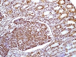
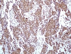
- Immunohistochemical analysis of HMGB1 in a section of human small intestinal cancer tissue. Samples were incubated in HMGB1 monoclonal antibody (Product # MA5-16264) using a dilution of 5 µg/mL. Intense nuclear immunopositivity of HMGB1 was observed in cancer cells and the cells of tumor stroma as well as the adjacent normal crypts. Some glandular cells in the crypts developed bother nuclear and cytoplasmic staining.

- Submitted by
- Invitrogen Antibodies (provider)
- Main image
- Experimental details
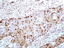
- Immunohistochemical analysis of HMGB1 in a tissue section of human esophageal squamous cell carcinoma (SCC). Samples were incubated in HMGB1 monoclonal antibody (Product # MA5-16264) using a dilution of 5 µg/mL. Strong nuclear along with weak cytoplasmic immunopositivity for HMGB1 was observed in SCC cells and the tumor stroma cells developed relatively weak staining for this target.

- Submitted by
- Invitrogen Antibodies (provider)
- Main image
- Experimental details
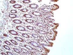
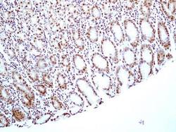
- Immunohistochemical analysis of HMGB1 in a tissue section of normal human colon. Samples were incubated in HMGB1 monoclonal antibody (Product # MA5-16264) using a dilution of 5 µg/mL. Almost all the cells of colons mucosal layer showed nuclear positivity for HMGB1 but the cells of the columnar epithelial cells on the absorptive surface developed cytoplasmic staining also.

- Submitted by
- Invitrogen Antibodies (provider)
- Main image
- Experimental details
- Immunohistochemical analysis of HMGB1 in a tissue section of human bladder transitional cell carcinoma (TCC) / urothelial cell carcinoma (UCC). Samples were incubated in HMGB1 monoclonal antibody (Product # MA5-16264) using a dilution of 5 µg/mL. Almost all the cancer cells and the cells of tumor stroma developed strong/specific nuclear HMGB1 immunopositivity.

- Submitted by
- Invitrogen Antibodies (provider)
- Main image
- Experimental details
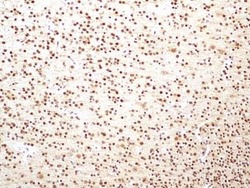
- Immunohistochemical analysis of HMGB1 in a tissue section of normal human brain. Samples were incubated in HMGB1 monoclonal antibody (Product # MA5-16264) using a dilution of 5 µg/mL. The representative image shows an overall strong nuclear HMGB1 immunopositivity with weak to negligible cytoplasmic staining in brain cells.

- Submitted by
- Invitrogen Antibodies (provider)
- Main image
- Experimental details
- Immunohistochemical analysis of HMGB1 in a section of normal human stomach tissue. Samples were incubated in HMGB1 monoclonal antibody (Product # MA5-16264) using a dilution of 5 µg/mL. The cells of the glandular stomach showed specific nuclear expression for HMGB1.

Supportive validation
- Submitted by
- Invitrogen Antibodies (provider)
- Main image

- Experimental details
- Flow cytometry of HMGB1 in Human Jurkat cells. Samples were incubated in HMGB1 monoclonal (Product # MA5-16264) using a dilution of 0.5 µg. Antibody (red) and 0.5 µg of isotype control antibody (green).
