Explore
 Validate
 Learn
Learn Western blot
Antibody data
- Antibody Data
- Antigen structure
- References [2]
- Comments [0]
- Validations
- Western blot [9]
- Immunocytochemistry [4]
- Immunohistochemistry [4]
- Other assay [1]
Submit
Validation data
Reference
Comment
Report error
- Product number
- PA5-27378 - Provider product page

- Provider
- Invitrogen Antibodies
- Product name
- HMGB1 Polyclonal Antibody
- Antibody type
- Polyclonal
- Antigen
- Recombinant protein fragment
- Description
- Recommended positive controls: 293T, A431, HeLa, HepG2, NIH-3T3, JC, BCL-1, C2C12, Raw264.7, PC-12, HMGB1-transfected 293T. Predicted reactivity: Mouse (100%), Rat (100%), Xenopus laevis (93%), Dog (100%), Pig (100%), Rabbit (100%), Chicken (94%), Rhesus Monkey (100%), Bovine (100%). Store product as a concentrated solution. Centrifuge briefly prior to opening the vial.
- Reactivity
- Human, Mouse, Rat, Porcine
- Host
- Rabbit
- Isotype
- IgG
- Vial size
- 100 µL
- Concentration
- 0.43 mg/mL
- Storage
- Store at 4°C short term. For long term storage, store at -20°C, avoiding freeze/thaw cycles.
Submitted references Inhibition of the Receptor for Advanced Glycation End Products Enhances the Cytotoxic Effect of Gemcitabine in Murine Pancreatic Tumors.
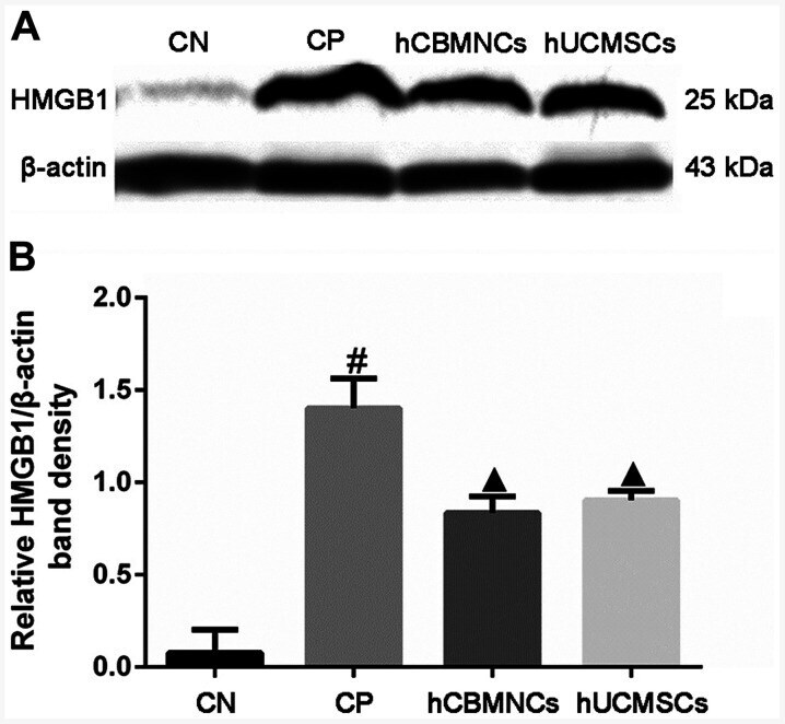
Human umbilical cord-derived mesenchymal stem cells and human cord blood mononuclear cells protect against cisplatin-induced acute kidney injury in rat models.
Swami P, O'Connell KA, Thiyagarajan S, Crawford A, Patil P, Radhakrishnan P, Shin S, Caffrey TC, Grunkemeyer J, Neville T, Vetter SW, Hollingsworth MA, Leclerc E
Biomolecules 2021 Apr 1;11(4)
Biomolecules 2021 Apr 1;11(4)
Human umbilical cord-derived mesenchymal stem cells and human cord blood mononuclear cells protect against cisplatin-induced acute kidney injury in rat models.
Xu Q, Yan P, Duan XJ, Wu X, Chen XJ, Luo M, Peng JC, Feng LX, Liu J, Zhong HL, Cheng W, Zou QY, Duan SB
Experimental and therapeutic medicine 2020 Dec;20(6):145
Experimental and therapeutic medicine 2020 Dec;20(6):145
No comments: Submit comment
Supportive validation
- Submitted by
- Invitrogen Antibodies (provider)
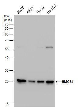
- Main image

- Experimental details
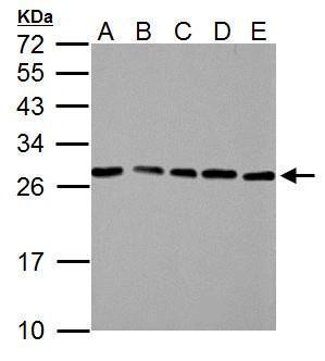
- Western blot analysis of HMGB1 using Various whole cell extracts (30 µg). Samples were loaded onto a 12% SDS-PAGE gel and probed with a HMGB1 polyclonal antibody (Product # PA5-27378) at a dilution of 1:3000.
- Submitted by
- Invitrogen Antibodies (provider)
- Main image

- Experimental details
- Western blot analysis of HMGB1 using A) 30 µg 293T whole cell lysate (B) 30 µg A431 whole cell lysate and C) 30 µg A375 whole cell lysate. Samples were loaded onto a 12% SDS-PAGE gel and probed with a HMGB1 polyclonal antibody (Product # PA5-27378) at a dilution of 1:3000.

- Submitted by
- Invitrogen Antibodies (provider)
- Main image
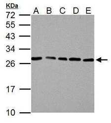
- Experimental details
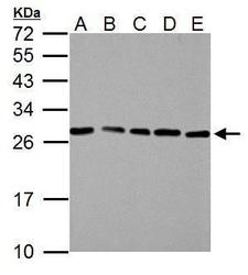
- HMGB1 Polyclonal Antibody detects HMGB1 protein by western blot analysis. A. 30 µg NIH-3T3 whole cell lysate/extract. B. 30 µg JC whole cell lysate/extract. C. 30 µg BCL-1 whole cell lysate/extract. D. 30 µg C2C12 whole cell lysate/extract. E. 30 µg Raw264.7 whole cell lysate/extract.12% SDS-PAGE. HMGB1 Polyclonal Antibody (Product # PA5-27378) dilution: 1:3,000. The HRP-conjugated anti-rabbit IgG antibody was used to detect the primary antibody.

- Submitted by
- Invitrogen Antibodies (provider)
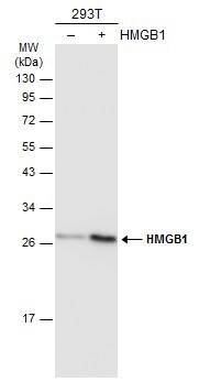
- Main image

- Experimental details
- Western Blot analysis of HMGB1 was performed by separating 30 µg of non-transfected (–) and transfected (+) 293T whole cell extracts by 12% SDS-PAGE. Proteins were transferred to a membrane and probed with a HMGB1 Polyclonal Antibody (Product # PA5-27378) at a dilution of 1:5000. The HRP-conjugated anti-rabbit IgG antibody was used to detect the primary antibody.
- Submitted by
- Invitrogen Antibodies (provider)
- Main image

- Experimental details
- Western Blot using HMGB1 Polyclonal Antibody (Product # PA5-27378). Various whole cell extracts (30 µg) were separated by 12% SDS-PAGE, and the membrane was blotted with HMGB1 Polyclonal Antibody (Product # PA5-27378) diluted at 1:3,000. The HRP-conjugated anti-rabbit IgG antibody was used to detect the primary antibody.

- Submitted by
- Invitrogen Antibodies (provider)
- Main image
- Experimental details
- HMGB1 Polyclonal Antibody detects HMGB1 protein by western blot analysis. A. 30 µg NIH-3T3 whole cell lysate/extract. B. 30 µg JC whole cell lysate/extract. C. 30 µg BCL-1 whole cell lysate/extract. D. 30 µg C2C12 whole cell lysate/extract. E. 30 µg Raw264.7 whole cell lysate/extract.12% SDS-PAGE. HMGB1 Polyclonal Antibody (Product # PA5-27378) dilution: 1:3,000. The HRP-conjugated anti-rabbit IgG antibody was used to detect the primary antibody.
- Submitted by
- Invitrogen Antibodies (provider)
- Main image

- Experimental details
- Western Blot analysis of HMGB1 was performed by separating 30 µg of various whole cell extracts by 12% SDS-PAGE. Proteins were transferred to a membrane and probed with a HMGB1 Polyclonal Antibody (Product # PA5-27378) at a dilution of 1:3000 and a HRP-conjugated anti-rabbit IgG secondary antibody.

- Submitted by
- Invitrogen Antibodies (provider)
- Main image
- Experimental details
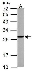
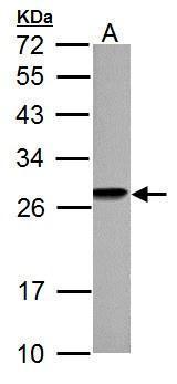
- HMGB1 Polyclonal Antibody detects HMGB1 protein by western blot analysis. A. 30 µg PC-12 whole cell lysate/extract.12% SDS-PAGE. HMGB1 Polyclonal Antibody (Product # PA5-27378) dilution: 1:3,000. The HRP-conjugated anti-rabbit IgG antibody was used to detect the primary antibody.
- Submitted by
- Invitrogen Antibodies (provider)
- Main image

- Experimental details
- Knockout of HMGB1 was achieved by CRISPR-Cas9 genome editing using LentiArray™ Lentiviral sgRNA (Product # A32042, Assay ID CRISPR784665_LV) and LentiArray Cas9 Lentivirus (Product # A32064). Western blot analysis of HMGB1 was performed by loading 30 µg of HeLa Wild Type (Lane 1), HeLa Cas9 (Lane 2) andHeLa HMGB1 KO (Lane 3) whole cell extracts. The samples were electrophoresed using NuPAGE™ Novex™ 4-12% Bis-Tris Protein Gel (Product # NP0322BOX). Resolved proteins were then transferred onto a nitrocellulose membrane (Product # IB23001) by iBlot® 2 Dry Blotting System (Product # IB21001). The blot was probed with Anti-HMGB1 Polyclonal Antibody (Product # PA5-27378, 1:5,000 dilution) and Goat anti-Rabbit IgG (H+L) Superclonal™ Recombinant Secondary Antibody, HRP (Product # A27036, 1:5,000 dilution) using the iBright FL 1000 (Product # A32752). Chemiluminescent detection was performed using Novex® ECL Chemiluminescent Substrate Reagent Kit (Product # WP20005). Loss of signal upon CRISPR mediated knockout (KO) using the LentiArray™ CRISPR product line confirms that antibody is specific to HMGB1. Uncharacterized band was observed in all the samples at 35 kDa.

Supportive validation
- Submitted by
- Invitrogen Antibodies (provider)
- Main image

- Experimental details
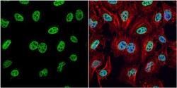
- Immunofluorescent analysis of HMGB1 showing staining in the nucleus of SK-N-SH cells. SK-N-SH cells were fixed in 4% paraformaldehyde at RT for 15 min and stained using a HMGB1 polyclonal antibody (Product # PA5-27378) diluted at 1:500. Blue: Hoechst 33342 staining. Scale bar = 10µm.

- Submitted by
- Invitrogen Antibodies (provider)
- Main image
- Experimental details
- Immunocytochemistry-Immunofluorescence analysis of HMGB1 was performed in HeLa cells fixed in 4% paraformaldehyde at RT for 15 min. Green: HMGB1 Polyclonal Antibody (Product # PA5-27378) diluted at 1:1000. Red: phalloidin, a cytoskeleton marker. Blue: Hoechst 33342 staining.

- Submitted by
- Invitrogen Antibodies (provider)
- Main image

- Experimental details
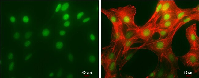
- Immunocytochemistry-Immunofluorescence analysis of HMGB1 was performed in NIH/3T3 cells fixed in 4% paraformaldehyde at RT for 15 min. Green: HMGB1 Polyclonal Antibody (Product # PA5-27378) diluted at 1:500. Red: phalloidin, a cytoskeleton marker. Scale bar = 10 µm.
- Submitted by
- Invitrogen Antibodies (provider)
- Main image
- Experimental details
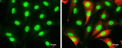
- Immunocytochemistry-Immunofluorescence analysis of HMGB1 was performed in SK-N-SH cells fixed in 4% paraformaldehyde at RT for 15 min. Green: HMGB1 Polyclonal Antibody (Product # PA5-27378) diluted at 1:1000. Red: beta Tubulin 3/ Tuj1, a cytoskeleton marker. Scale bar = 10 µm.

Supportive validation
- Submitted by
- Invitrogen Antibodies (provider)
- Main image

- Experimental details
- Immunohistochemistry (Paraffin) analysis of HMGB1 was performed in paraffin-embedded mouse esophagus tissue using HMGB1 Polyclonal Antibody (Product # PA5-27378) at a dilution of 1:1000.

- Submitted by
- Invitrogen Antibodies (provider)
- Main image
- Experimental details
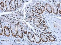
- HMGB1 Polyclonal Antibody detects HMGB1 protein at nucleus on mouse colon by immunohistochemical analysis. Sample: Paraffin-embedded mouse colon. HMGB1 Polyclonal Antibody (Product # PA5-27378) dilution: 1:1,000. Antigen Retrieval: EDTA based buffer, pH 8.0, 15 min.

- Submitted by
- Invitrogen Antibodies (provider)
- Main image

- Experimental details
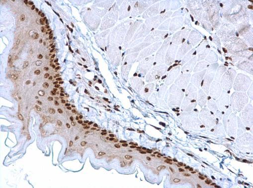
- HMGB1 Polyclonal Antibody detects HMGB1 protein at nucleus on mouse esophagus by immunohistochemical analysis. Sample: Paraffin-embedded mouse esophagus. HMGB1 Polyclonal Antibody (Product # PA5-27378) dilution: 1:1,000. Antigen Retrieval: EDTA based buffer, pH 8.0, 15 min.
- Submitted by
- Invitrogen Antibodies (provider)
- Main image
- Experimental details
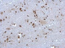
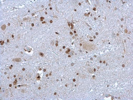
- HMGB1 Polyclonal Antibody detects HMGB1 protein at nucleus on rat brain stem by immunohistochemical analysis. Sample: Paraffin-embedded rat brain stem. HMGB1 Polyclonal Antibody (Product # PA5-27378) dilution: 1:1,000. Antigen Retrieval: EDTA based buffer, pH 8.0, 15 min.
Supportive validation
- Submitted by
- Invitrogen Antibodies (provider)
- Main image

- Experimental details
- Figure 6 Protein expression of HMGB1 in the renal tissues of each group. (A) Protein levels of HMBG1 were measured using western blotting. (B) Protein bands were quantified using Tanon 5200 Multi Image Analysis software and relative HMGB1/beta-actin band densities were measured. Data are presented as the mean +- SD (n=6/group). # P