Explore
 Validate
 Learn
Learn Western blot
 Immunocytochemistry
Antibody data
- Antibody Data
- Antigen structure
- References [8]
- Comments [0]
- Validations
- Western blot [1]
- Flow cytometry [1]
- Chromatin Immunoprecipitation [1]
Submit
Validation data
Reference
Comment
Report error
- Product number
- AF3639 - Provider product page

- Provider
- Novus Biologicals
- Product name
- Goat Polyclonal Snail Antibody
- Antibody type
- Polyclonal
- Description
- Antigen Affinity-purified. Detects human Snail in direct ELISAs and Western blots.
- Reactivity
- Human
- Host
- Goat
- Conjugate
- Unconjugated
- Isotype
- IgG
- Vial size
- 100 ug
- Concentration
- LYOPH
- Storage
- Use a manual defrost freezer and avoid repeated freeze-thaw cycles. 12 months from date of receipt, -20 to -70 degreesC as supplied. 1 month, 2 to 8 degreesC under sterile conditions after reconstitution. 6 months, -20 to -70 degreesC under sterile conditions after reconstitution.
Submitted references The ubiquitin-specific protease USP17 prevents cellular senescence by stabilizing the methyltransferase SET8 and transcriptionally repressing p21.
Ginsenoside Rk1 Induces Apoptosis in Neuroblastoma Cells Through Loss of Mitochondrial Membrane Potential and Activation of Caspases.
The Oncogenic Activity of miR-29b-1-5p Induces the Epithelial-Mesenchymal Transition in Oral Squamous Cell Carcinoma.
MDM2 promotes epithelial-mesenchymal transition and metastasis of ovarian cancer SKOV3 cells.
Pro-invasive properties of Snail1 are regulated by sumoylation in response to TGFβ stimulation in cancer.
Pan-cancer EMT-signature identifies RBM47 down-regulation during colorectal cancer progression.
Epithelial-mesenchymal transitioned circulating tumor cells capture for detecting tumor progression.
SNAI1 expression and the mesenchymal phenotype: an immunohistochemical study performed on 46 cases of oral squamous cell carcinoma.
Fukuura K, Inoue Y, Miyajima C, Watanabe S, Tokugawa M, Morishita D, Ohoka N, Komada M, Hayashi H
The Journal of biological chemistry 2019 Nov 1;294(44):16429-16439
The Journal of biological chemistry 2019 Nov 1;294(44):16429-16439
Ginsenoside Rk1 Induces Apoptosis in Neuroblastoma Cells Through Loss of Mitochondrial Membrane Potential and Activation of Caspases.
Oh JM, Lee J, Im WT, Chun S
International journal of molecular sciences 2019 Mar 11;20(5)
International journal of molecular sciences 2019 Mar 11;20(5)
The Oncogenic Activity of miR-29b-1-5p Induces the Epithelial-Mesenchymal Transition in Oral Squamous Cell Carcinoma.
Kurihara-Shimomura M, Sasahira T, Shimomura H, Nakashima C, Kirita T
Journal of clinical medicine 2019 Feb 24;8(2)
Journal of clinical medicine 2019 Feb 24;8(2)
MDM2 promotes epithelial-mesenchymal transition and metastasis of ovarian cancer SKOV3 cells.
Chen Y, Wang DD, Wu YP, Su D, Zhou TY, Gai RH, Fu YY, Zheng L, He QJ, Zhu H, Yang B
British journal of cancer 2017 Oct 10;117(8):1192-1201
British journal of cancer 2017 Oct 10;117(8):1192-1201
Pro-invasive properties of Snail1 are regulated by sumoylation in response to TGFβ stimulation in cancer.
Gudey SK, Sundar R, Heldin CH, Bergh A, Landström M
Oncotarget 2017 Nov 17;8(58):97703-97726
Oncotarget 2017 Nov 17;8(58):97703-97726
Pan-cancer EMT-signature identifies RBM47 down-regulation during colorectal cancer progression.
Rokavec M, Kaller M, Horst D, Hermeking H
Scientific reports 2017 Jul 5;7(1):4687
Scientific reports 2017 Jul 5;7(1):4687
Epithelial-mesenchymal transitioned circulating tumor cells capture for detecting tumor progression.
Satelli A, Mitra A, Brownlee Z, Xia X, Bellister S, Overman MJ, Kopetz S, Ellis LM, Meng QH, Li S
Clinical cancer research : an official journal of the American Association for Cancer Research 2015 Feb 15;21(4):899-906
Clinical cancer research : an official journal of the American Association for Cancer Research 2015 Feb 15;21(4):899-906
SNAI1 expression and the mesenchymal phenotype: an immunohistochemical study performed on 46 cases of oral squamous cell carcinoma.
Schwock J, Bradley G, Ho JC, Perez-Ordonez B, Hedley DW, Irish JC, Geddie WR
BMC clinical pathology 2010 Feb 5;10:1
BMC clinical pathology 2010 Feb 5;10:1
No comments: Submit comment
Supportive validation
- Submitted by
- Novus Biologicals (provider)
- Main image
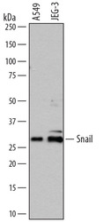
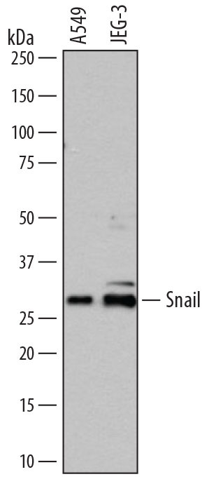
- Experimental details
- Detection of Human Snail by Western Blot. Western blot shows lysates of A549 human lung carcinoma cell line and JEG-3 human epithelial choriocarcinoma cell line. PVDF membrane was probed with 0.5 µg/mL of Goat Anti-Human Snail Antigen Affinity-purified Polyclonal Antibody (Catalog # AF3639) followed by HRP-conjugated Anti-Goat IgG Secondary Antibody (Catalog # HAF109). A specific band was detected for Snail at approximately 29 kDa (as indicated). This experiment was conducted under reducing conditions and using Immunoblot Buffer Group 1.
Supportive validation
- Submitted by
- Novus Biologicals (provider)
- Main image
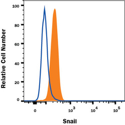
- Experimental details
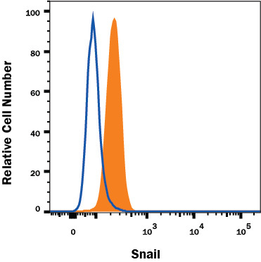
- Detection of Snail in A549 Human Cell Line by Flow Cytometry. A549 human lung carcinoma cell line was stained with Goat Anti-Human Snail Antigen Affinity-purified Polyclonal Antibody (Catalog # AF3639, filled histogram) or isotype control antibody (Catalog # AB-108-C, open histogram), followed by Fluorescein-conjugated Anti-Goat IgG Secondary Antibody (Catalog # F0109). To facilitate intracellular staining, cells were fixed and permeabilized with FlowX FoxP3 Fixation & Permeabilization Buffer Kit (Catalog # FC012). View our protocol for Staining Intracellular Molecules.
Supportive validation
- Submitted by
- Novus Biologicals (provider)
- Main image
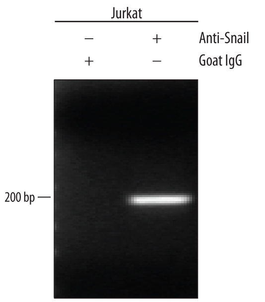
- Experimental details
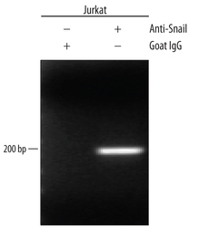
- Detection of Snail-regulated Genes by Chromatin Immunoprecipitation. Jurkat human acute T cell leukemia cell line treated with 50 ng/mL PMA and 200 ng/mL calcium ionomycin for 30 minutes was fixed using formaldehyde, resuspended in lysis buffer, and sonicated to shear chromatin. Snail/DNA complexes were immunoprecipitated using 5 μg Goat Anti-Human Snail Antigen Affinity-purified Polyclonal Antibody (Catalog # AF3639) or control antibody (Catalog # AB-108-C) for 15 minutes in an ultrasonic bath, followed by Biotinylated Anti-Goat IgG Secondary Antibody (Catalog # BAF109). Immunocomplexes were captured using 50 μL of MagCellect Streptavidin Ferrofluid (Catalog # MAG999) and DNA was purified using chelating resin solution. The E-Cadherin promoter was detected by standard PCR.