Explore
 Validate
 Learn
Learn Western blot
 Immunocytochemistry
Antibody data
- Antibody Data
- Antigen structure
- References [0]
- Comments [0]
- Validations
- Immunocytochemistry [6]
- Immunohistochemistry [5]
- Flow cytometry [1]
Submit
Validation data
Reference
Comment
Report error
- Product number
- MA5-34709 - Provider product page

- Provider
- Invitrogen Antibodies
- Product name
- EEF1A1 Recombinant Rabbit Monoclonal Antibody (JB44-13)
- Antibody type
- Monoclonal
- Antigen
- Synthetic peptide
- Description
- Positive Control: Rat brain tissue, mouse skeletal muscle tissue lysate, rat skin tissue lysate, Daudi, mouse cerebellum tissue lysate, HepG2, HUVEC, SH-SY-5Y, rat skeletal muscle tissue, human tonsil tissue, human liver cancer tissue, human kidney tissue, mouse smooth muscle tissue, THP-1.
- Reactivity
- Human
- Host
- Rabbit
- Isotype
- IgG
- Antibody clone number
- JB44-13
- Vial size
- 100 μL
- Concentration
- 1 mg/mL
- Storage
- -20°C, Avoid Freeze/Thaw Cycles, store in dark
No comments: Submit comment
Supportive validation
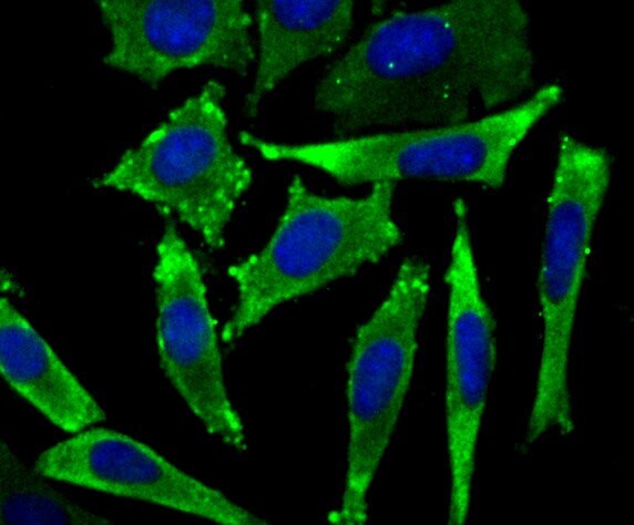
- Submitted by
- Invitrogen Antibodies (provider)
- Main image

- Experimental details
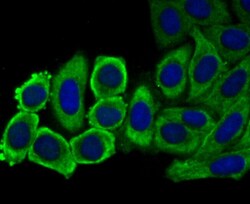
- Immunofluorescent analysis of eEF1A1 in SH-SY-5Y cells (green). Samples were fixed in paraformaldehyde and permeabilised with 0.25% Triton X100/PBS, incubated with eEF1A1 monoclonal antibody (Product # MA5-34709), followed by DAPI (blue).

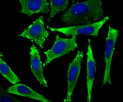
- Submitted by
- Invitrogen Antibodies (provider)
- Main image
- Experimental details
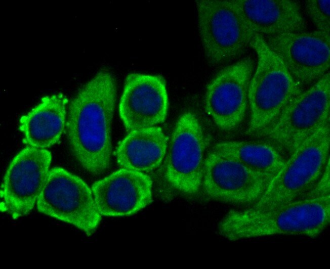
- Immunofluorescent analysis of eEF1A1 in HUVEC cells (green). Samples were fixed in paraformaldehyde and permeabilised with 0.25% Triton X100/PBS, incubated with eEF1A1 monoclonal antibody (Product # MA5-34709), followed by DAPI (blue).

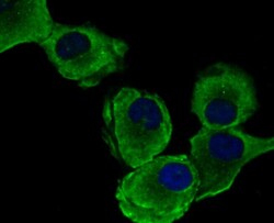
- Submitted by
- Invitrogen Antibodies (provider)
- Main image

- Experimental details
- Immunofluorescent analysis of eEF1A1 in HepG2 cells (green). Samples were fixed in paraformaldehyde and permeabilised with 0.25% Triton X100/PBS, incubated with eEF1A1 monoclonal antibody (Product # MA5-34709), followed by DAPI (blue).

- Submitted by
- Invitrogen Antibodies (provider)
- Main image

- Experimental details
- Immunofluorescent analysis of eEF1A1 in HUVEC cells (green). Samples were fixed in paraformaldehyde and permeabilised with 0.25% Triton X100/PBS, incubated with eEF1A1 monoclonal antibody (Product # MA5-34709), followed by DAPI (blue).

- Submitted by
- Invitrogen Antibodies (provider)
- Main image
- Experimental details
- Immunofluorescent analysis of eEF1A1 in SH-SY-5Y cells (green). Samples were fixed in paraformaldehyde and permeabilised with 0.25% Triton X100/PBS, incubated with eEF1A1 monoclonal antibody (Product # MA5-34709), followed by DAPI (blue).
- Submitted by
- Invitrogen Antibodies (provider)
- Main image
- Experimental details
- Immunofluorescent analysis of eEF1A1 in HepG2 cells (green). Samples were fixed in paraformaldehyde and permeabilised with 0.25% Triton X100/PBS, incubated with eEF1A1 monoclonal antibody (Product # MA5-34709), followed by DAPI (blue).
Supportive validation
- Submitted by
- Invitrogen Antibodies (provider)
- Main image

- Experimental details
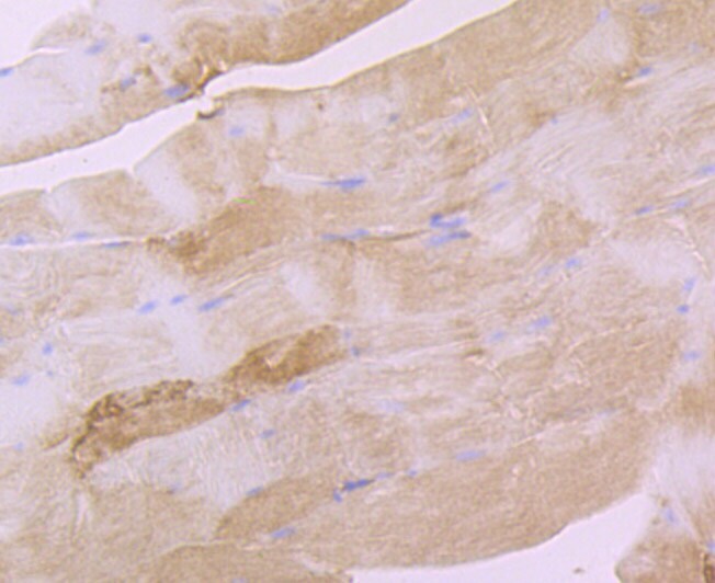
- Immunohistochemistry analysis of eEF1A1 in paraffin-embedded mouse smooth muscle tissue. Samples were incubated with eEF1A1 monoclonal antibody (Product # MA5-34709), and followed by hematoxylin.
- Submitted by
- Invitrogen Antibodies (provider)
- Main image
- Experimental details
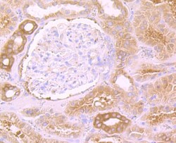
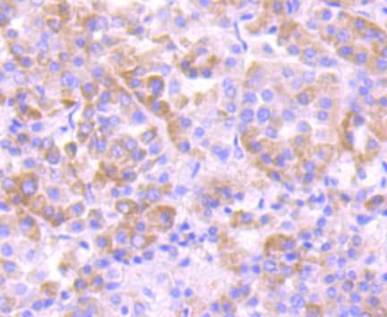
- Immunohistochemistry analysis of eEF1A1 in paraffin-embedded human kidney tissue. Samples were incubated with eEF1A1 monoclonal antibody (Product # MA5-34709), and followed by hematoxylin.

- Submitted by
- Invitrogen Antibodies (provider)
- Main image

- Experimental details
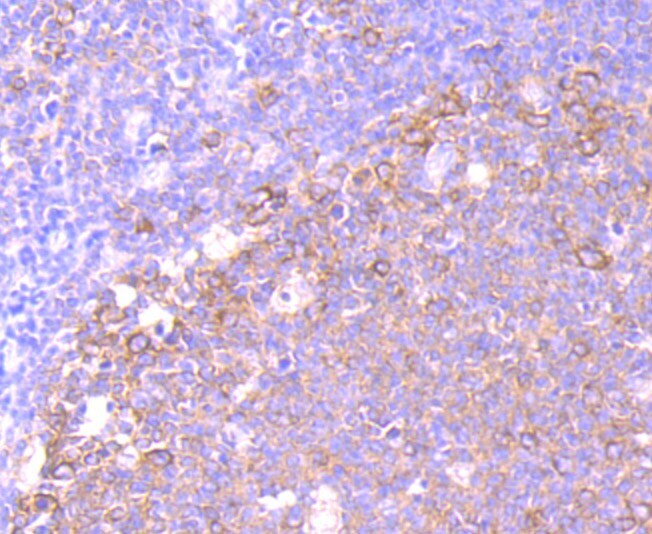
- Immunohistochemistry analysis of eEF1A1 in paraffin-embedded human tonsil tissue. Samples were incubated with eEF1A1 monoclonal antibody (Product # MA5-34709), and followed by hematoxylin.
- Submitted by
- Invitrogen Antibodies (provider)
- Main image

- Experimental details
- Immunohistochemistry analysis of eEF1A1 in paraffin-embedded rat skeletal muscle tissue. Samples were incubated with eEF1A1 monoclonal antibody (Product # MA5-34709), and followed by hematoxylin.

- Submitted by
- Invitrogen Antibodies (provider)
- Main image
- Experimental details
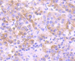
- Immunohistochemistry analysis of eEF1A1 in paraffin-embedded human liver cancer tissue. Samples were incubated with eEF1A1 monoclonal antibody (Product # MA5-34709), and followed by hematoxylin.
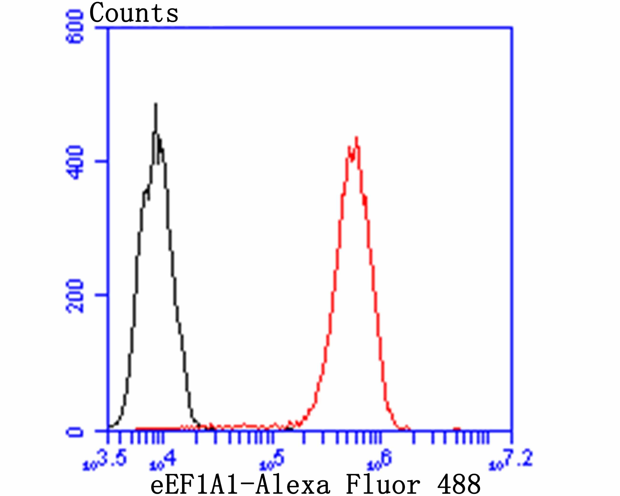
Supportive validation
- Submitted by
- Invitrogen Antibodies (provider)
- Main image
- Experimental details
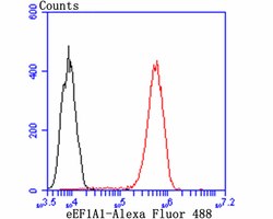
- Flow cytometry of eEF1A1 in THP-1 cells (red) compared with an unlabelled control (cells without incubation with primary antibody; black). Samples were incubated with eEF1A1 monoclonal antibody (Product # MA5-34709) at a dilution of 1:100, followed by Alexa Fluor 488-conjugated goat anti-rabbit IgG.