Explore
 Validate
 Learn
Learn Western blot
Antibody data
- Antibody Data
- Antigen structure
- References [1]
- Comments [0]
- Validations
- Western blot [4]
- Immunocytochemistry [1]
- Other assay [2]
Submit
Validation data
Reference
Comment
Report error
- Product number
- PA5-17213 - Provider product page

- Provider
- Invitrogen Antibodies
- Product name
- EEF1A1 Polyclonal Antibody
- Antibody type
- Polyclonal
- Antigen
- Synthetic peptide
- Description
- It is not recommended to aliquot this antibody.
- Reactivity
- Human, Mouse, Rat
- Host
- Rabbit
- Isotype
- IgG
- Vial size
- 100 µL
- Concentration
- 50.5 µg/mL
- Storage
- -20°C
Submitted references The Eukaryotic Elongation Factor 1 Alpha (eEF1α) from the Parasite Leishmania infantum Is Modified with the Immunomodulatory Substituent Phosphorylcholine (PC).
Timm T, Annoscia G, Klein J, Lochnit G
Molecules (Basel, Switzerland) 2017 Nov 29;22(12)
Molecules (Basel, Switzerland) 2017 Nov 29;22(12)
No comments: Submit comment
Supportive validation
- Submitted by
- Invitrogen Antibodies (provider)
- Main image

- Experimental details
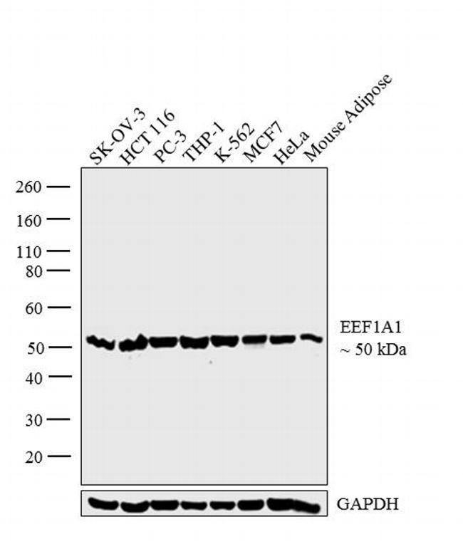
- Western blot analysis was performed on whole cell extracts (30 µg lysate) of SK-OV-3 (Lane 1), HCT 116 (Lane 2), PC-3 (Lane 3), THP-1 (Lane 4), K-562 (Lane 5), MCF7 (Lane 6), HeLa (Lane 7) and tissue extract (30 µg lysate) of Mouse Adipose (Lane 8). The blot was probed with Anti-EEF1A1 Polyclonal Antibody (Product # PA5-17213, 1:3000 dilution) and detected by chemiluminescence using Goat anti-Rabbit IgG (H+L) Superclonal™ Secondary Antibody, HRP conjugate (Product # A27036, 0.25 µg/ml, 1:4000 dilution). A 50 kDa band corresponding to EEF1A1 was observed across the cell lines and tissue tested.
- Submitted by
- Invitrogen Antibodies (provider)
- Main image

- Experimental details
- Western blot analysis was performed on whole cell extracts (30 µg lysate) of SK-OV-3 (Lane 1), HCT 116 (Lane 2), PC-3 (Lane 3), THP-1 (Lane 4), K-562 (Lane 5), MCF7 (Lane 6), HeLa (Lane 7) and tissue extract (30 µg lysate) of Mouse Adipose (Lane 8). The blot was probed with Anti-EEF1A1 Polyclonal Antibody (Product # PA5-17213, 1:3000 dilution) and detected by chemiluminescence using Goat anti-Rabbit IgG (H+L) Superclonal™ Secondary Antibody, HRP conjugate (Product # A27036, 0.25 µg/ml, 1:4000 dilution). A 50 kDa band corresponding to EEF1A1 was observed across the cell lines and tissue tested.

- Submitted by
- Invitrogen Antibodies (provider)
- Main image
- Experimental details
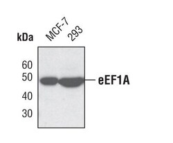
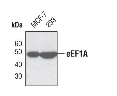
- Western blot analysis of eEF1A in extracts from MCF-7 and 293 cells using eEF1A polyclonal antibody (Product # PA5-17213).
- Submitted by
- Invitrogen Antibodies (provider)
- Main image

- Experimental details
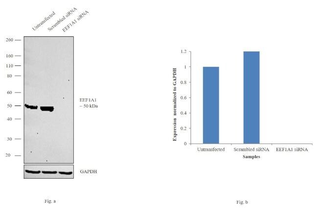
- Knockdown of EEF1A1 was achieved by transfecting MCF7 with EEF1A1 specific siRNAs (Silencer® select Product # s4477, s4478). Western blot analysis (Fig. a) was performed using whole cell extracts from the EEF1A1 knockdown cells (lane 3), non-specific scrambled siRNA transfected cells (lane 2) and untransfected cells (lane 1). The blot was probed with EEF1A1 Polyclonal Antibody (Product # PA5-17213, 1:1000 dilution) and Goat anti-Rabbit IgG (H+L) Superclonal™ Secondary Antibody, HRP conjugate (Product # A27036, 0.25 µg/mL, 1:4000 dilution). Densitometric analysis of this western blot is shown in histogram (Fig. b). Decrease in signal upon siRNA mediated knock down confirms that antibody is specific to EEF1A1.
Supportive validation
- Submitted by
- Invitrogen Antibodies (provider)
- Main image
- Experimental details
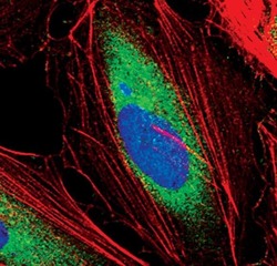
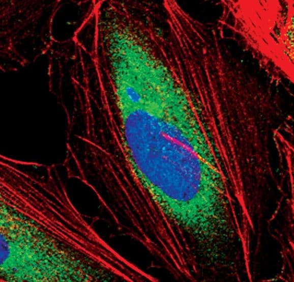
- Immunofluorescent analysis of eEF1A in HeLa cells using an EEF1A polyclonal antibody (Product # PA5-17213) (green). Actin filaments are labeled with a fluorescent red phalloidin. DNA is labeled using a fluorescent blue dye.
Supportive validation
- Submitted by
- Invitrogen Antibodies (provider)
- Main image
- Experimental details
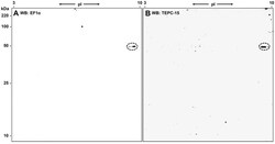
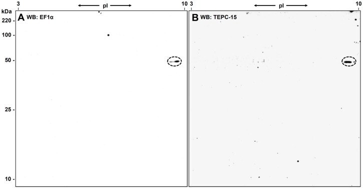
- Figure 4 2D-Western blot analyses with eEF1alpha- and PC-specific antibodies. ( A ) ECL of a Western blot probed with PA5-17213, an eEF1alpha-specific antibody. The elongation factor is detected at its expected molecular weight (49.5 kDa) but at a slightly higher pI (9.5 compared to an expected pI of 9.03). There is also a second spot of the same size but at lower pI detectable, representing another protein species of eEF1alpha ( B ). Both spots of eEF1alpha shown in ( A ) are recognized by the PC-specific antibody TEPC-15. Note that this Leishmania were cultivated for ten days but display no difference in PC-substitution compared to the ones cultivated only for three days.
- Submitted by
- Invitrogen Antibodies (provider)
- Main image
- Experimental details
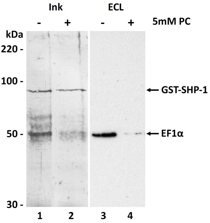
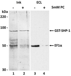
- Figure 7 Interaction of L. infantum EF1alpha with human SHP-1. Leishmania EF1alpha co-purifies with human GST-SHP-1 as detected in a Western blot by the eEF1alpha-specific antibody (lane 3 ). This interaction is almost completely abolished in presence of 5 mM phosphorylcholine (lane 4 ). Lanes 1 and 2 show an Ink-staining of the proteins co-eluting with the GST-tagged phosphatase SHP-1 directly on the PVDF membrane. Whereas the SHP-1 (~90 kDa) is visible in both lanes at similar amounts, a band corresponding to EF1alpha (~50 kDa) is much stronger in lane 1 than in lane 2, were PC interrupts their interaction.