Explore
 Validate
 Learn
Learn Western blot
 ELISA
ELISAAntibody data
- Antibody Data
- Antigen structure
- References [7]
- Comments [0]
- Validations
- Western blot [1]
- Immunohistochemistry [2]
Submit
Validation data
Reference
Comment
Report error
- Product number
- 15638-1-AP - Provider product page

- Provider
- Proteintech Group
- Proper citation
- Proteintech Cat#15638-1-AP, RRID:AB_2158168
- Product name
- OPA3 antibody
- Antibody type
- Polyclonal
- Description
- OPA3 antibody (Cat. #15638-1-AP) is a rabbit polyclonal antibody that shows reactivity with human, mouse, rat and has been validated for the following applications: IF, IHC, IP, WB,ELISA.
- Reactivity
- Human, Mouse, Rat
- Host
- Rabbit
- Conjugate
- Unconjugated
- Isotype
- IgG
- Vial size
- 20ul, 150ul
Submitted references OPA3 inhibits the cGAS-STING pathway mediated by mtDNA stress to promote colorectal cancer progression.
Hydrogen sulfide alleviates mitochondrial damage and ferroptosis by regulating OPA3-NFS1 axis in doxorubicin-induced cardiotoxicity.
MYB proto-oncogene like 2 promotes hepatocellular carcinoma growth and glycolysis via binding to the Optic atrophy 3 promoter and activating its expression.
Oncogenic K-ras Induces Mitochondrial OPA3 Expression to Promote Energy Metabolism in Pancreatic Cancer Cells.
Disrupted mitochondrial function in the Opa3L122P mouse model for Costeff Syndrome impairs skeletal integrity.
Mitochondrial localization and ocular expression of mutant Opa3 in a mouse model of 3-methylglutaconicaciduria type III.
A nonsense mutation in the optic atrophy 3 gene (OPA3) causes dilated cardiomyopathy in Red Holstein cattle.
Yin Y, Ma Z, Yuan S, Xu K, Wang X
In vitro cellular & developmental biology. Animal 2025 Feb;61(2):165-177
In vitro cellular & developmental biology. Animal 2025 Feb;61(2):165-177
Hydrogen sulfide alleviates mitochondrial damage and ferroptosis by regulating OPA3-NFS1 axis in doxorubicin-induced cardiotoxicity.
Wang Y, Ying X, Wang Y, Zou Z, Yuan A, Xiao Z, Geng N, Qiao Z, Li W, Lu X, Pu J
Cellular signalling 2023 Jul;107:110655
Cellular signalling 2023 Jul;107:110655
MYB proto-oncogene like 2 promotes hepatocellular carcinoma growth and glycolysis via binding to the Optic atrophy 3 promoter and activating its expression.
Liu M, Du Q, Mao G, Dai N, Zhang F
Bioengineered 2022 Mar;13(3):5344-5356
Bioengineered 2022 Mar;13(3):5344-5356
Oncogenic K-ras Induces Mitochondrial OPA3 Expression to Promote Energy Metabolism in Pancreatic Cancer Cells.
Meng N, Glorieux C, Zhang Y, Liang L, Zeng P, Lu W, Huang P
Cancers 2019 Dec 25;12(1)
Cancers 2019 Dec 25;12(1)
Disrupted mitochondrial function in the Opa3L122P mouse model for Costeff Syndrome impairs skeletal integrity.
Navein AE, Cooke EJ, Davies JR, Smith TG, Wells LH, Ohazama A, Healy C, Sharpe PT, Evans SL, Evans BA, Votruba M, Wells T
Human molecular genetics 2016 Jun 15;25(12):2404-2416
Human molecular genetics 2016 Jun 15;25(12):2404-2416
Mitochondrial localization and ocular expression of mutant Opa3 in a mouse model of 3-methylglutaconicaciduria type III.
Powell KA, Davies JR, Taylor E, Wride MA, Votruba M
Investigative ophthalmology & visual science 2011 Jun 21;52(7):4369-80
Investigative ophthalmology & visual science 2011 Jun 21;52(7):4369-80
A nonsense mutation in the optic atrophy 3 gene (OPA3) causes dilated cardiomyopathy in Red Holstein cattle.
Owczarek-Lipska M, Plattet P, Zipperle L, Drögemüller C, Posthaus H, Dolf G, Braunschweig MH
Genomics 2011 Jan;97(1):51-7
Genomics 2011 Jan;97(1):51-7
No comments: Submit comment
Supportive validation
- Submitted by
- Proteintech Group (provider)
- Main image

- Experimental details
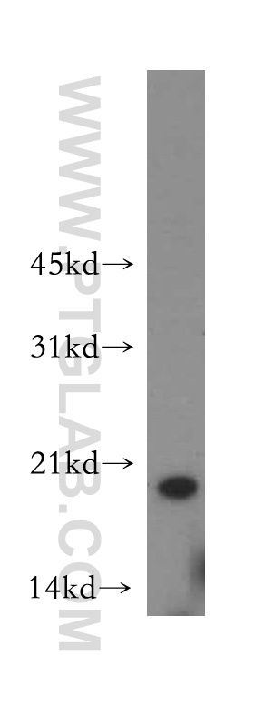
- HeLa cells were subjected to SDS PAGE followed by western blot with 15638-1-AP(OPA3 antibody) at dilution of 1:400
- Sample type
- cell line
Supportive validation
- Submitted by
- Proteintech Group (provider)
- Main image
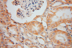
- Experimental details
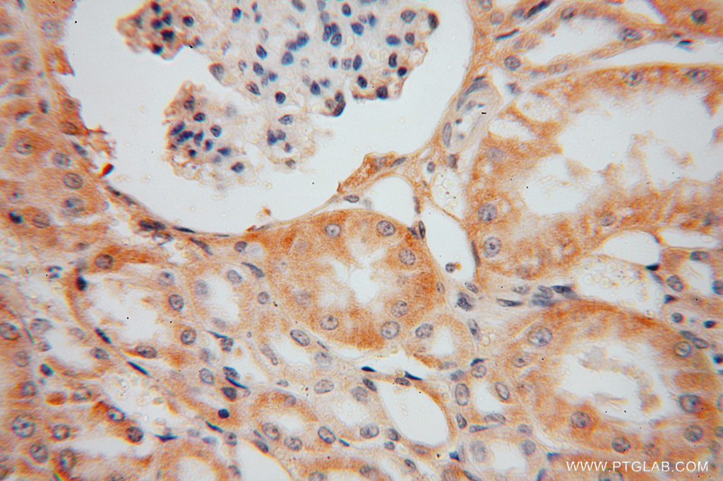
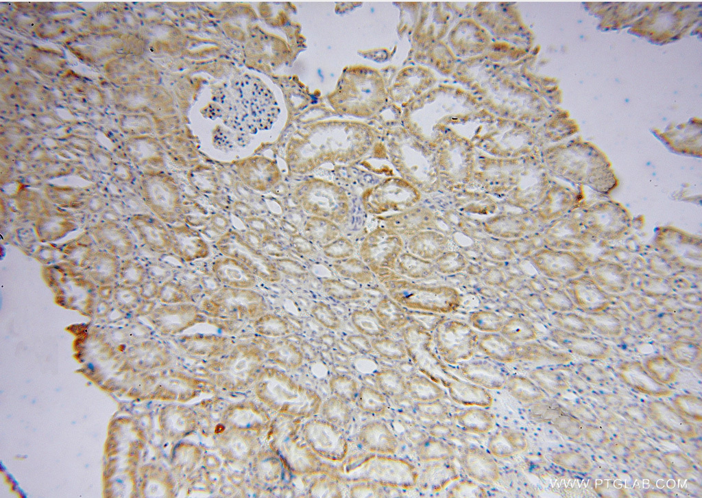
- Immunohistochemical of paraffin-embedded human kidney using 15638-1-AP(OPA3 antibody) at dilution of 1:50 (under 10x lens)
- Sample type
- tissue
- Submitted by
- Proteintech Group (provider)
- Main image
- Experimental details
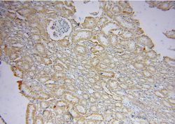
- The OPA3 antibody from Proteintech is a rabbit polyclonal antibody to a recombinant protein of human OPA3. This antibody recognizes human, mouse, rat antigen. The OPA3 antibody has been validated for the following applications: ELISA, WB, IHC analysis.