Explore
 Validate
 Learn
Learn Western blot
Antibody data
- Antibody Data
- Antigen structure
- References [0]
- Comments [0]
- Validations
- Western blot [2]
- Immunocytochemistry [1]
- Immunohistochemistry [1]
Submit
Validation data
Reference
Comment
Report error
- Product number
- TA324484 - Provider product page

- Provider
- OriGene
- Product name
- Rabbit polyclonal LSD1 Antibody (C-term)
- Antibody type
- Polyclonal
- Description
- Rabbit polyclonal LSD1 Antibody (C-term)
- Host
- Rabbit
- Conjugate
- Unconjugated
- Epitope
- KDM1A
- Isotype
- IgG
- Antibody clone number
- NULL
- Vial size
- 400 µl
- Concentration
- 2.0 mg/ml
No comments: Submit comment
Supportive validation
- Submitted by
- OriGene (provider)
- Main image

- Experimental details
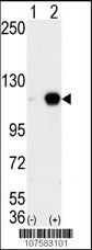
- Western blot analysis of AOF2 (arrow) using LSD1 Antibody (C-term) (Cat.#TA324484). 293 cell lysates (2 ug/lane) either nontransfected (Lane 1) or transiently transfected with the AOF2 gene (Lane 2) (Origene Technologies).
- Validation comment
- WB
- Submitted by
- OriGene (provider)
- Main image

- Experimental details
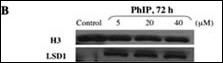
- Western immunoblot. Nuclear extracts of control and PhIP-treated HMEC. Proteins were transferred onto polyvinylidene difluoride and blotted with anti-LSD1 antibody. Nuclear LSD1 protein levels increased in carcinogen-treated HMEC compared with control HMEC.
- Validation comment
- WB
Supportive validation
- Submitted by
- OriGene (provider)
- Main image

- Experimental details
- IF image of Hela cell stained with hLSD1-Y712(Cat#TA324484). Hela cells were incubated with hLSD1 primary antibody (1:25, 1 h at 37?). For secondary antibody, Alexa Fluor? 488 conjugated donkey anti-rabbit antibody (green) was used (1:400).Cytoplasmic actin was counterstained with Alexa Fluor? 555 (red) conjugated Phalloidin (7 units/ml). Nuclei were counterstained with DAPI (blue) . hLSD1 immunoreactivity is localized to nucleus significantly.
- Validation comment
- IF

Supportive validation
- Submitted by
- OriGene (provider)
- Main image

- Experimental details
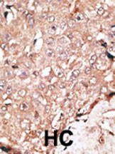
- Formalin-fixed and paraffin-embedded human cancer tissue reacted with the primary antibody, which was peroxidase-conjugated to the secondary antibody, followed by DAB staining. This data demonstrates the use of this antibody for immunohistochemistry; clinical relevance has not been evaluated. BC = breast carcinoma; HC = hepatocarcinoma.
- Validation comment
- IHC