Explore
 Validate
 Learn
Learn Western blot
 Immunocytochemistry
Antibody data
- Antibody Data
- Antigen structure
- References [7]
- Comments [0]
- Validations
- Western blot [1]
Submit
Validation data
Reference
Comment
Report error
- Product number
- PA1780 - Provider product page

- Provider
- Boster Biological Technology
- Product name
- Anti-VDAC/Porin/VDAC1 Antibody
- Antibody type
- Polyclonal
- Description
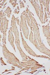
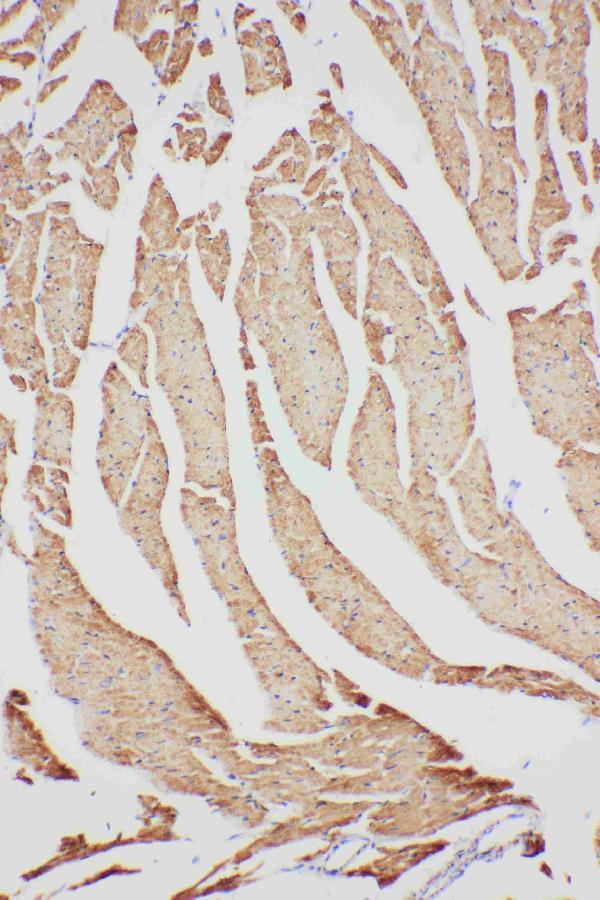
- Polyclonal antibody for PORIN/VDAC1 detection. Host: Rabbit.Size: 100μg/vial. Tested applications: IHC-P. Reactive species: Human. PORIN/VDAC1 information: Molecular Weight: 30773 MW; Subcellular Localization: Mitochondrion outer membrane . Cell membrane ; Tissue Specificity: Heart, liver and skeletal muscle.
- Reactivity
- Human, Mouse, Rat
- Host
- Rabbit
- Vial size
- 100μg/vial
- Concentration
- Add 0.2ml of distilled water will yield a concentration of 500ug/ml.
- Storage
- At -20°C for one year. After reconstitution, at 4°C for one month. It can also be aliquoted and stored frozen at -20°C for a longer time. Avoid repeated freezing and thawing.
- Handling
- Add 0.2ml of distilled water will yield a concentration of 500ug/ml.
Submitted references TERT Promoter Revertant Mutation Inhibits Melanoma Growth through Intrinsic Apoptosis.
Deficiency of β-carotene oxygenase 2 induces mitochondrial fragmentation and activates the STING-IRF3 pathway in the mouse hypothalamus.
Fetal Programming and Sexual Dimorphism of Mitochondrial Protein Expression and Activity of Hearts of Prenatally Hypoxic Guinea Pig Offspring.
Sexual dimorphism of mitochondrial function in the hypoxic guinea pig placenta.
Rotenone induces nephrotoxicity in rats: oxidative damage and apoptosis.
Novel chemiluminescent Western blot blocking and antibody incubation solution for enhanced antibody-antigen interaction and increased specificity.
Lack of β, β-carotene-9', 10'-oxygenase 2 leads to hepatic mitochondrial dysfunction and cellular oxidative stress in mice.
Wang Y, Chen Y, Li C, Xiao Z, Yuan H, Zhang Y, Pang D, Tang X, Li M, Ouyang H
Biology 2022 Jan 14;11(1)
Biology 2022 Jan 14;11(1)
Deficiency of β-carotene oxygenase 2 induces mitochondrial fragmentation and activates the STING-IRF3 pathway in the mouse hypothalamus.
Wu L, Guo X, Wong SY, Lu P, Hartson SD, Medeiros DM, Wang W, Clarke SL, Lucas EA, Smith BJ, Chowanadisai W, Lin D
The Journal of nutritional biochemistry 2021 Feb;88:108542
The Journal of nutritional biochemistry 2021 Feb;88:108542
Fetal Programming and Sexual Dimorphism of Mitochondrial Protein Expression and Activity of Hearts of Prenatally Hypoxic Guinea Pig Offspring.
Thompson LP, Song H, Polster BM
Oxidative medicine and cellular longevity 2019;2019:7210249
Oxidative medicine and cellular longevity 2019;2019:7210249
Sexual dimorphism of mitochondrial function in the hypoxic guinea pig placenta.
Song H, Telugu BP, Thompson LP
Biology of reproduction 2019 Jan 1;100(1):208-216
Biology of reproduction 2019 Jan 1;100(1):208-216
Rotenone induces nephrotoxicity in rats: oxidative damage and apoptosis.
Jiang XW, Qiao L, Feng XX, Liu L, Wei QW, Wang XW, Yu WH
Toxicology mechanisms and methods 2017 Sep;27(7):528-536
Toxicology mechanisms and methods 2017 Sep;27(7):528-536
Novel chemiluminescent Western blot blocking and antibody incubation solution for enhanced antibody-antigen interaction and increased specificity.
Schwartz K, Bochkariov D
Electrophoresis 2017 Oct;38(20):2631-2637
Electrophoresis 2017 Oct;38(20):2631-2637
Lack of β, β-carotene-9', 10'-oxygenase 2 leads to hepatic mitochondrial dysfunction and cellular oxidative stress in mice.
Wu L, Guo X, Hartson SD, Davis MA, He H, Medeiros DM, Wang W, Clarke SL, Lucas EA, Smith BJ, von Lintig J, Lin D
Molecular nutrition & food research 2017 May;61(5)
Molecular nutrition & food research 2017 May;61(5)
No comments: Submit comment
Supportive validation
- Submitted by
- Boster Biological Technology (provider)
- Main image
- Experimental details
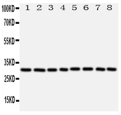
- Anti-VDAC/Porin antibody, PA1780, Western blottingAll lanes: Anti VDAC/Porin (PA1780) at 0.5ug/ml Lane 1: Rat Skeletal Muscle Tissue Lysate at 50ug Lane 2: Rat Heart Tissue Lysate at 50ug Lane 3: Rat Liver Tissue Lysate at 50ug Lane 4: HELA Whole Cell Lysate at 40ug Lane 5: A431 Whole Cell Lysate at 40ug Lane 6: A549 Whole Cell Lysate at 40ug Lane 7: SMMC Whole Cell Lysate at 40ug Lane 8: HT1080 Whole Cell Lysate at 40ug Predicted bind size: 31KD Observed bind size: 31KD
- Additional image