Explore
 Validate
 Learn
Learn Western blot
Antibody data
- Antibody Data
- Antigen structure
- References [1]
- Comments [0]
- Validations
- Western blot [2]
- Immunohistochemistry [2]
- Other assay [1]
Submit
Validation data
Reference
Comment
Report error
- Product number
- PA5-68530 - Provider product page

- Provider
- Invitrogen Antibodies
- Product name
- SLC26A3 Polyclonal Antibody
- Antibody type
- Polyclonal
- Antigen
- Synthetic peptide
- Description
- This target displays homology in the following species: Cow: 100%; Dog: 79%; Guinea Pig: 79%; Human: 100%; Mouse: 93%; Pig: 100%; Rabbit: 86%; Rat: 93%; Sheep: 100%
- Reactivity
- Human
- Host
- Rabbit
- Isotype
- IgG
- Vial size
- 100 μL
- Concentration
- 0.5 mg/mL
- Storage
- -20°C, Avoid Freeze/Thaw Cycles
Submitted references Inhibition of intestinal villus cell Na/K-ATPase mediates altered glucose and NaCl absorption in obesity-associated diabetes and hypertension.
Palaniappan B, Arthur S, Sundaram VL, Butts M, Sundaram S, Mani K, Singh S, Nepal N, Sundaram U
FASEB journal : official publication of the Federation of American Societies for Experimental Biology 2019 Aug;33(8):9323-9333
FASEB journal : official publication of the Federation of American Societies for Experimental Biology 2019 Aug;33(8):9323-9333
No comments: Submit comment
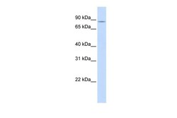
Supportive validation
- Submitted by
- Invitrogen Antibodies (provider)
- Main image
- Experimental details
- Western blot analysis of SLC26A3 on human pancreas tissue lysate. The sample was probed with a SLC26A3 polyclonal antibody (Product # PA5-68530) using a primary antibody dilution of 0.2-1.0 µg/mL.

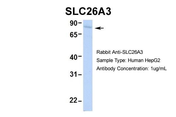
- Submitted by
- Invitrogen Antibodies (provider)
- Main image
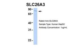
- Experimental details
- Western blot analysis of SLC26A3 on HepG2 cells. The sample was probed with a SLC26A3 polyclonal antibody (Product # PA5-68530) using a primary antibody dilution of 1.0 µg/mL.
Supportive validation
- Submitted by
- Invitrogen Antibodies (provider)
- Main image
- Experimental details
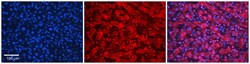
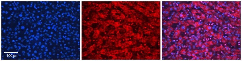
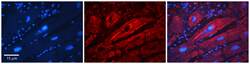
- Immunohistochemical analysis of SLC26A3 in paraffin-embedded human adult liver tissue. Sample was probed with a SLC26A3 polyclonal antibody (Product # PA5-68530) at a dilution of 1:600. Detection was performed with a donkey anti-Rabbit Cy2/3 secondary antibody at a dilution of 1:200. Images were taken at 20x magnification with an exposure time of 0.5-2.0 seconds.
- Submitted by
- Invitrogen Antibodies (provider)
- Main image
- Experimental details
- Immunohistochemical analysis of SLC26A3 in paraffin-embedded human adult liver tissue. Sample was probed with a SLC26A3 polyclonal antibody (Product # PA5-68530) at a dilution of 1:600. Detection was performed with a donkey anti-Rabbit Cy2/3 secondary antibody at a dilution of 1:200. Images were taken at 20x magnification with an exposure time of 0.5-2.0 seconds.

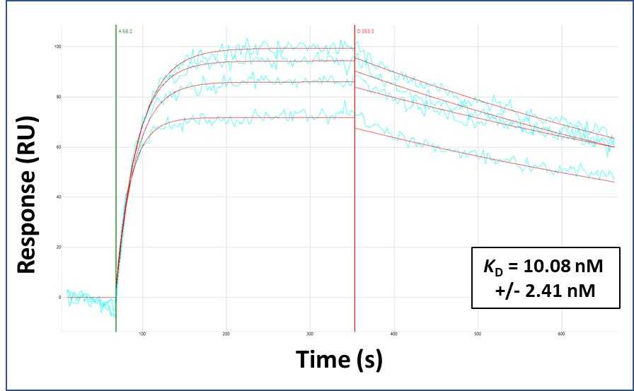
Supportive validation
- Submitted by
- Invitrogen Antibodies (provider)
- Main image
- Experimental details
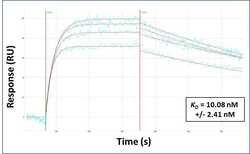
- Surface Plasmon Resonance of SLC26A3 polyclonal antibody (Product # PA5-68530). Purified polyclonal antibodies were immobilized on a Protein A/G coated Carterra LSA sensor chip at concentrations of 5, and 50 µg/mL in duplicate. Antibodies on the surface were exposed to interaction with peptides sequentially via microfluidic controlled flow at 333 nm peptide concentration for kinetic characterization of the binders for affinity and specificity, followed by curve fitting using the Kinetics software. Kd determinations for both concentrations were averaged and results and standard deviation are shown.