Explore
 Validate
 Learn
Learn Immunocytochemistry
Antibody data
- Antibody Data
- Antigen structure
- References [3]
- Comments [0]
- Validations
- Immunocytochemistry [1]
Submit
Validation data
Reference
Comment
Report error
- Product number
- HPA019887 - Provider product page

- Provider
- Atlas Antibodies
- Proper citation
- Atlas Antibodies Cat#HPA019887, RRID:AB_1855080
- Product name
- Anti-PCNT
- Antibody type
- Polyclonal
- Description
- Polyclonal Antibody against Human PCNT, Gene description: pericentrin, Alternative Gene Names: KEN, KIAA0402, PCN, PCNT2, PCNTB, SCKL4, Validated applications: ICC, Uniprot ID: O95613, Storage: Store at +4°C for short term storage. Long time storage is recommended at -20°C.
- Reactivity
- Human
- Host
- Rabbit
- Conjugate
- Unconjugated
- Isotype
- IgG
- Vial size
- 100 µl
- Concentration
- 0.05 mg/ml
- Storage
- Store at +4°C for short term storage. Long time storage is recommended at -20°C.
- Handling
- The antibody solution should be gently mixed before use.
Submitted references Cilia defects upon loss of WDR4 are linked to proteasomal hyperactivity and ubiquitin shortage
Dyslexia Candidate Gene and Ciliary Gene Expression Dynamics During Human Neuronal Differentiation.
Resting cells rely on the DNA helicase component MCM2 to build cilia
Burkhalter M, Stiff T, Maerz L, Casar Tena T, Wiese H, Gerhards J, Sailer S, Vu L, Duong Phu M, Donow C, Alupei M, Iben S, Groth M, Wiese S, Church J, Jeggo P, Philipp M
Cell Death & Disease 2024;15(9)
Cell Death & Disease 2024;15(9)
Dyslexia Candidate Gene and Ciliary Gene Expression Dynamics During Human Neuronal Differentiation.
Bieder A, Yoshihara M, Katayama S, Krjutškov K, Falk A, Kere J, Tapia-Páez I
Molecular neurobiology 2020 Jul;57(7):2944-2958
Molecular neurobiology 2020 Jul;57(7):2944-2958
Resting cells rely on the DNA helicase component MCM2 to build cilia
Casar Tena T, Maerz L, Szafranski K, Groth M, Blätte T, Donow C, Matysik S, Walther P, Jeggo P, Burkhalter M, Philipp M
Nucleic Acids Research 2019;47(1):134-151
Nucleic Acids Research 2019;47(1):134-151
No comments: Submit comment
Supportive validation
- Submitted by
- Atlas Antibodies (provider)
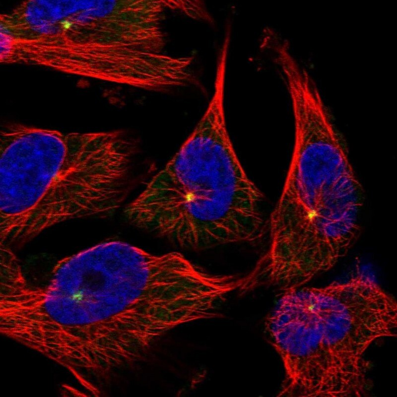
- Main image
- Experimental details
- Immunofluorescent staining of human cell line U-251 MG shows localization to centrosome.
- Sample type
- Human