 Explore
 Validate
 Learn
LearnPA5-53498
antibody from Invitrogen Antibodies
Targeting: PCNT
KEN, KIAA0402, PCN, PCNT2, PCNTB, SCKL4
 Immunocytochemistry
Antibody data
- Antibody Data
- Antigen structure
- References [0]
- Comments [0]
- Validations
- Immunocytochemistry [4]
- Immunohistochemistry [5]
Submit
Validation data
Reference
Comment
Report error
- Product number
- PA5-53498 - Provider product page

- Provider
- Invitrogen Antibodies
- Product name
- Pericentrin Polyclonal Antibody
- Antibody type
- Polyclonal
- Antigen
- Recombinant full-length protein
- Description
- Immunogen sequence: MEDLQNQFQK ELAEQRAELE KIFQDKNQAE RALRNLESHH QAAIEKLRED LQSEHGRCLE DLEFKFKESE KEKQLELENL QASYEDLKAQ SQEEIRRLWS QLDSARTSRQ ELSELHEQLL ARTSRVEDLE QLKQREKTQH ESELE
- Concentration
- 0.4 mg/mL
No comments: Submit comment
Supportive validation
- Submitted by
- Invitrogen Antibodies (provider)
- Main image
- Experimental details
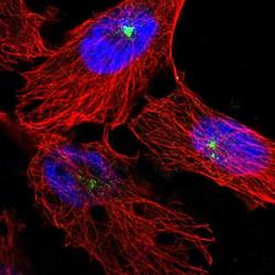
- Immunofluorescent staining of Pericentrin in human cell line U-251 MG shows positivity in centrosome. Samples were probed using a Pericentrin Polyclonal Antibody (Product # PA5-53498).
- Submitted by
- Invitrogen Antibodies (provider)
- Main image

- Experimental details
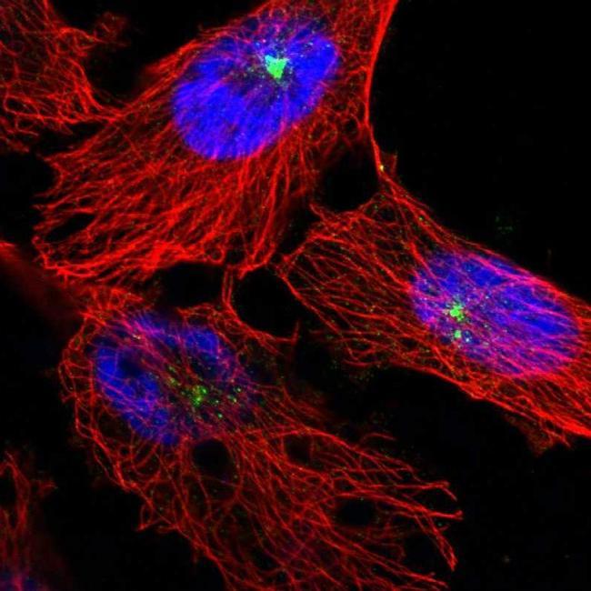
- Immunofluorescent staining of Pericentrin in human cell line A-431 using a Pericentrin Polyclonal Antibody (Product # PA5-53498) shows localization to centrosome.

- Submitted by
- Invitrogen Antibodies (provider)
- Main image

- Experimental details
- Immunofluorescence analysis of Pericentrin was performed using MCF7 cells. The cells were fixed with 4% paraformaldehyde for 10 minutes, permeabilized with 0.1% Triton™ X-100 for 15 minutes, and blocked with 1% BSA for 1 hour at room temperature. The cells were labeled with Pericentrin Rabbit Polyclonal Antibody (Product # PA5-53498) at 4µg/mL dilution in 0.1% BSA and incubated overnight at 4 degree and then labeled with Goat anti-Rabbit IgG (H+L) Superclonal™ Secondary Antibody, Alexa Fluor® 488 conjugate (Product # A27034) at a dilution of 1:2000 for 45 minutes at room temperature (Panel a: green). Nuclei (Panel b: blue) were stained with ProLong™ Diamond Antifade Mountant with DAPI (Product # P36962). F-actin (Panel c: red) was stained with Rhodamine Phalloidin (Product # R415, 1:300). Panel d represents the composite image showing centrosomal localization of Pericentrin. Panel e represents control cells with no primary antibody to assess background. The images were captured at 60X magnification. .

- Submitted by
- Invitrogen Antibodies (provider)
- Main image
- Experimental details
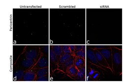
- KD of Pericentrin was achieved by transfecting MCF7 cells with Pericentrin specific siRNA (Silencer® select Product # s10136, s10137). Immunofluorescence analysis was performed on untransfected MCF7 cells (panel a,d), transfected with non-specific scrambled siRNA (panels b,e) and transfected with Pericentrin specific siRNA (panel c,f). Cells were fixed, permeabilized, and labelled with Pericentrin Rabbit Polyclonal Antibody (Product # PA5-53498, 4 µg/mL), followed by Goat anti-Rabbit IgG (H+L) Superclonal™ Secondary Antibody, Alexa Fluor® 488 conjugate (Product # A27034, 1:2000). Nuclei (blue) were stained using ProLong™ Diamond Antifade Mountant with DAPI (Product # P36962), and Rhodamine Phalloidin (Product # R415, 1:300) was used for cytoskeletal F-actin (red) staining. Reduction of specific signal was observed upon siRNA mediated KD (panel c,f) confirming specificity of the antibody to Pericentrin (green). The images were captured at 60X magnification..

Supportive validation
- Submitted by
- Invitrogen Antibodies (provider)
- Main image

- Experimental details
- Immunohistochemical staining of Pericentrin in human gallbladder using Pericentrin Polyclonal Antibody (Product # PA5-53498).

- Submitted by
- Invitrogen Antibodies (provider)
- Main image

- Experimental details
- Immunohistochemical staining of Pericentrin in human lymph node using Pericentrin Polyclonal Antibody (Product # PA5-53498).

- Submitted by
- Invitrogen Antibodies (provider)
- Main image

- Experimental details
- Immunohistochemical staining of Pericentrin in human testis using Pericentrin Polyclonal Antibody (Product # PA5-53498) shows high expression.

- Submitted by
- Invitrogen Antibodies (provider)
- Main image

- Experimental details
- Immunohistochemical staining of Pericentrin in human gallbladder, liver, lymph node and testis using Pericentrin Polyclonal Antibody (Product # PA5-53498) (A) shows similar protein distribution across tissues to an independent Pericentrin Polyclonal Antibody (B).

- Submitted by
- Invitrogen Antibodies (provider)
- Main image

- Experimental details
- Immunohistochemical staining of Pericentrin in human liver using Pericentrin Polyclonal Antibody (Product # PA5-53498) shows low expression as expected.
